
Search Count: 6
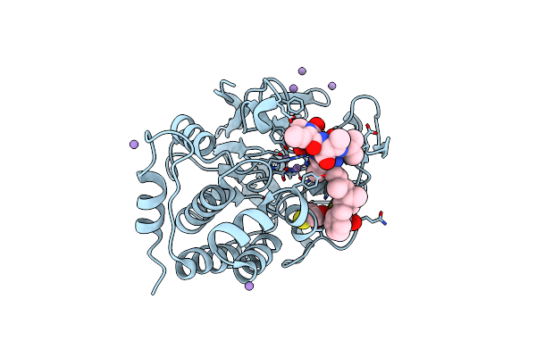 |
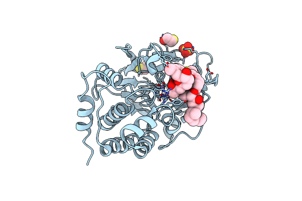
X-Ray Crystal Structure Of Protein Phosphatase-1 With The Marine Toxin Motuporin Bound
Organism: Homo sapiens, Marine sponge thenonella swinhoie grey
Method: X-RAY DIFFRACTION Resolution:2.10 Å Release Date: 2006-01-17 Classification: HYDROLASE/HYDROLASE INHIBITOR Ligands: BME, MN |
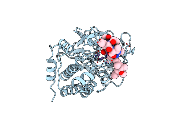 |
X-Ray Crystal Structure Of Dihydromicrocystin-La Bound To Protein Phosphatase-1
Organism: Homo sapiens, Microcystis aeruginosa
Method: X-RAY DIFFRACTION Resolution:2.30 Å Release Date: 2006-01-17 Classification: HYDROLASE/HYDROLASE INHIBITOR Ligands: MN |
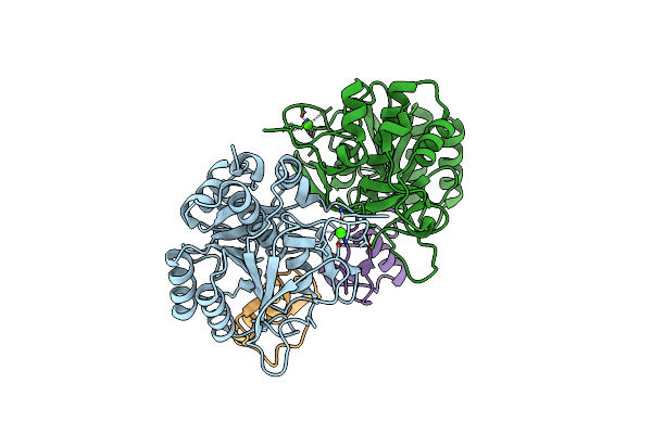 |
Organism: Meleagris gallopavo, Bacillus licheniformis
Method: X-RAY DIFFRACTION Resolution:1.55 Å Release Date: 2005-05-03 Classification: HYDROLASE Ligands: CA |
 |
Crystal Structure Of A Protein Phosphatase-1: Calcineurin Hybrid Bound To Okadaic Acid
Organism: Homo sapiens
Method: X-RAY DIFFRACTION Resolution:2.00 Å Release Date: 2004-08-17 Classification: HYDROLASE Ligands: MN, OKA, BME |
 |
The Crystal Structure Of 1D-Myo-Inositol 2-Acetamido-2-Deoxy-Alpha-D-Glucopyranoside Deacetylase (Mshb)
Organism: Mycobacterium tuberculosis
Method: X-RAY DIFFRACTION Resolution:1.70 Å Release Date: 2003-12-02 Classification: HYDROLASE Ligands: ZN, PE4 |
 |
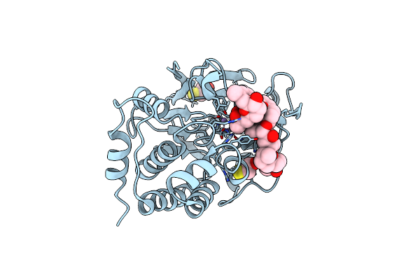
Crystal Structure Of The Tumor-Promoter Okadaic Acid Bound To Protein Phosphatase-1
Organism: Homo sapiens
Method: X-RAY DIFFRACTION Resolution:1.90 Å Release Date: 2001-08-15 Classification: HYDROLASE/TOXIN Ligands: MN, SO4, OKA, BME |