
Search Count: 245
 |
Organism: Homo sapiens
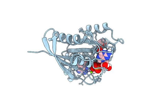
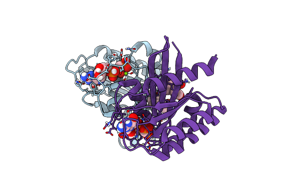
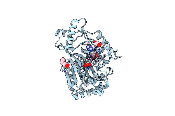
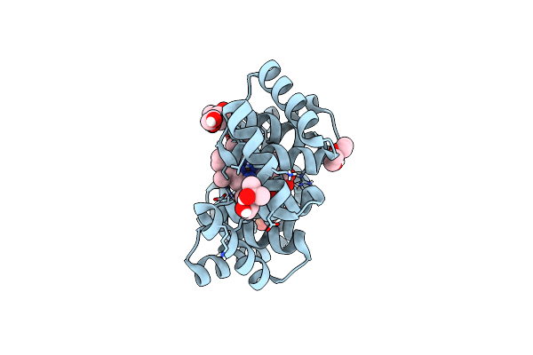
Method: X-RAY DIFFRACTION Release Date: 2025-08-06 Classification: HYDROLASE Ligands: GDP, MG, EDO, A1I1P |
 |
Organism: Homo sapiens
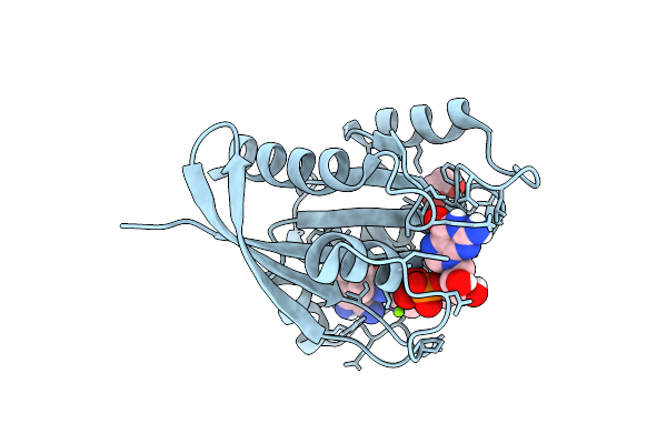
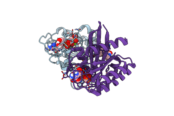
Method: X-RAY DIFFRACTION Release Date: 2025-08-06 Classification: HYDROLASE Ligands: GDP, MG, EDO, A1I1R |
 |
Organism: Homo sapiens
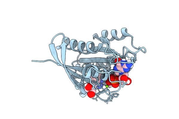
Method: X-RAY DIFFRACTION Release Date: 2025-08-06 Classification: HYDROLASE Ligands: GDP, MG, EDO, WYU |
 |
Organism: Homo sapiens
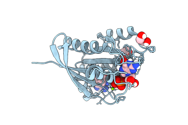
Method: X-RAY DIFFRACTION Release Date: 2025-08-06 Classification: HYDROLASE Ligands: GDP, MG, EDO, A1I1Z |
 |
Organism: Homo sapiens
Method: X-RAY DIFFRACTION Release Date: 2025-08-06 Classification: HYDROLASE Ligands: GDP, MG, EDO, VU6 |
 |
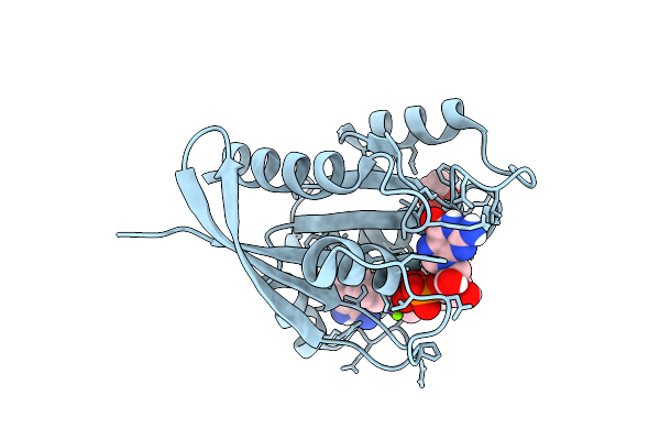
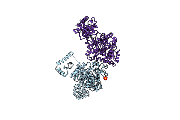
Spokes 12 And 13 Of The Human Gamma-Tubulin Ring Complex In Complex With Cdk5Rap2 And Docked Mzt2/Gcp2-Nhd Module
Organism: Homo sapiens
Method: ELECTRON MICROSCOPY Release Date: 2025-01-15 Classification: CELL CYCLE Ligands: GDP |
 |
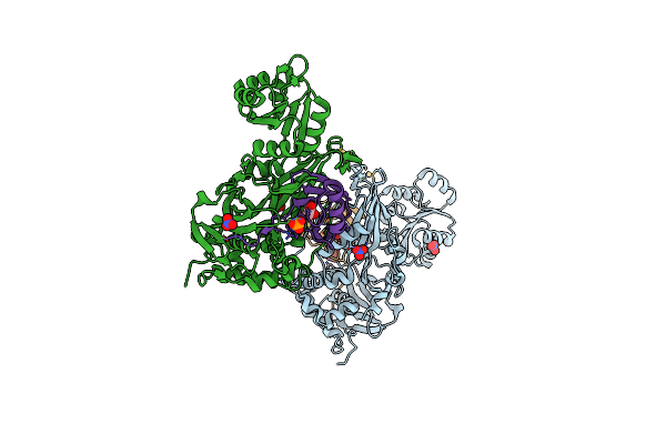
Candida Albicans Gamma-Tubulin Small Complex Within Ring-Like Higher Oligomer In Complex With Spc72 Cm1
Organism: Candida albicans
Method: ELECTRON MICROSCOPY Release Date: 2025-01-15 Classification: CELL CYCLE |
 |
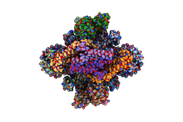
Full Gamma-Tubulin Ring Complex Composed Of The Candida Albicans Gamma-Tubulin Small Complex In Complex With Spc72 Cm1
Organism: Candida albicans
Method: ELECTRON MICROSCOPY Release Date: 2025-01-15 Classification: CELL CYCLE |
 |
Organism: Homo sapiens
Method: X-RAY DIFFRACTION Resolution:1.16 Å Release Date: 2024-10-02 Classification: ONCOPROTEIN Ligands: GCP, YLE, MG |
 |
Organism: Homo sapiens
Method: X-RAY DIFFRACTION Resolution:1.31 Å Release Date: 2024-10-02 Classification: ONCOPROTEIN Ligands: GCP, MG, YFJ |
 |
Organism: Homo sapiens
Method: X-RAY DIFFRACTION Resolution:1.25 Å Release Date: 2024-08-07 Classification: PROTEIN BINDING Ligands: A1IGX |
 |
Organism: Homo sapiens
Method: X-RAY DIFFRACTION Resolution:1.88 Å Release Date: 2024-08-07 Classification: PROTEIN BINDING Ligands: 5WX |
 |
Crystal Structure Of The L-Arginine Hydroxylase Vioc Mehis316, Bound To Fe(Ii), L-Arginine, And Succinate
Organism: Synthetic construct
Method: X-RAY DIFFRACTION Resolution:1.60 Å Release Date: 2024-07-31 Classification: OXIDOREDUCTASE Ligands: FE2, SIN, ARG, EDO, PEG |
 |
Organism: Homo sapiens
Method: X-RAY DIFFRACTION Resolution:1.74 Å Release Date: 2024-03-06 Classification: TRANSCRIPTION |
 |
Organism: Homo sapiens
Method: X-RAY DIFFRACTION Resolution:1.55 Å Release Date: 2024-03-06 Classification: TRANSCRIPTION Ligands: XS8 |
 |
Organism: Bacillus cereus vd045
Method: X-RAY DIFFRACTION Resolution:3.00 Å Release Date: 2023-11-22 Classification: DNA BINDING PROTEIN/Hydrolase Ligands: SO4 |
 |
Structure Of The Phage Immune Evasion Protein Gad1 Bound To The Gabija Gajab Complex
Organism: Bacillus cereus vd045, Bacillus phage phi3t
Method: ELECTRON MICROSCOPY Release Date: 2023-11-22 Classification: VIRAL PROTEIN |
 |
Crystal Structure Of Ubiquitin Specific Protease 11 (Usp11) In Complex With A Substrate Mimetic
Organism: Homo sapiens
Method: X-RAY DIFFRACTION Resolution:2.44 Å Release Date: 2023-10-18 Classification: HYDROLASE Ligands: CD, CL, NO3, PO4, GOL |
 |
Crystal Structure Of A Computationally Designed Heme Binding Protein, Dnhem1
Organism: Synthetic construct
Method: X-RAY DIFFRACTION Resolution:1.60 Å Release Date: 2023-07-05 Classification: BIOSYNTHETIC PROTEIN Ligands: IMD, HEM, MPD, PO4, PEG, EDO |
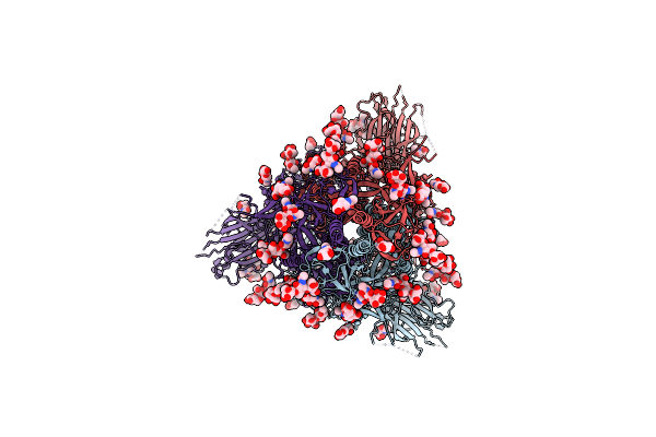 |
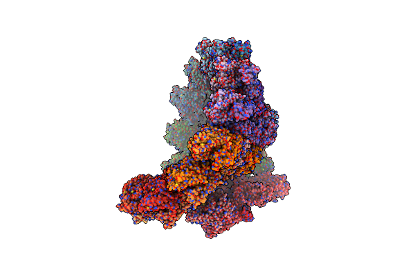
Organism: Severe acute respiratory syndrome coronavirus 2
Method: ELECTRON MICROSCOPY Release Date: 2023-06-07 Classification: VIRAL PROTEIN Ligands: NAG |