
Search Count: 15
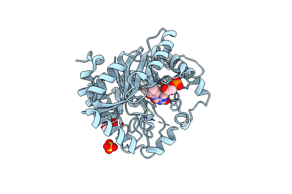 |
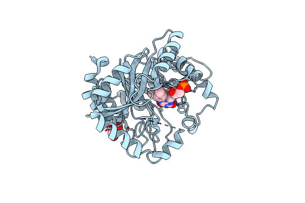
Crystal Structure Of Monooxygenase Ruta Complexed With Dioxygen Under 1.5 Mpa / 15 Bars Of Oxygen Pressure.
Organism: Escherichia coli (strain k12)
Method: X-RAY DIFFRACTION Resolution:1.80 Å Release Date: 2020-02-05 Classification: FLAVOPROTEIN Ligands: SO4, GOL, FMN, OXY |
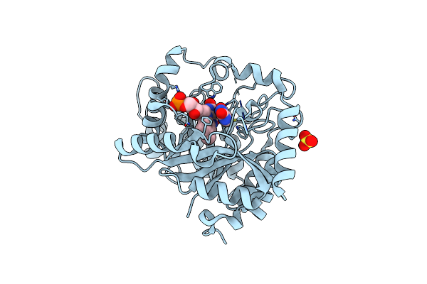 |
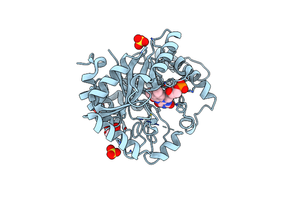
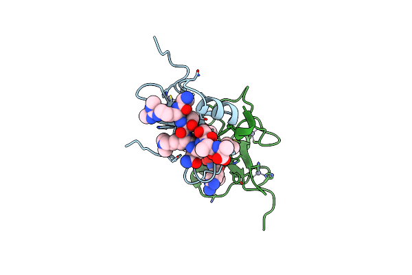
Crystal Structure Of Monooxygenase Ruta Complexed With Uracil Under Atmospheric Pressure.
Organism: Escherichia coli (strain k12)
Method: X-RAY DIFFRACTION Resolution:2.01 Å Release Date: 2020-02-05 Classification: FLAVOPROTEIN Ligands: FMN, URA, SO4 |
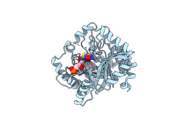 |
Organism: Escherichia coli (strain k12)
Method: X-RAY DIFFRACTION Resolution:2.00 Å Release Date: 2020-02-05 Classification: FLAVOPROTEIN Ligands: FMN, LDB |
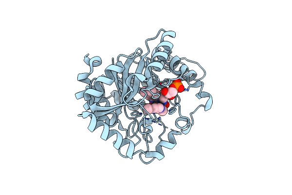 |
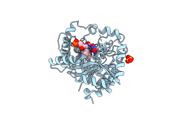
Crystal Structure Of Monooxygenase Ruta Complexed With 2,4-Dimethoxypyrimidine.
Organism: Escherichia coli (strain k12)
Method: X-RAY DIFFRACTION Resolution:2.50 Å Release Date: 2020-02-05 Classification: FLAVOPROTEIN Ligands: FMN, LD8 |
 |
Organism: Escherichia coli k-12
Method: X-RAY DIFFRACTION Resolution:2.20 Å Release Date: 2020-02-05 Classification: FLAVOPROTEIN Ligands: GOL, FMN |
 |
Crystal Structure Of Monooxygenase Ruta Complexed With Dioxygen Under 0.5 Mpa / 5 Bars Of Oxygen Pressure.
Organism: Escherichia coli k-12
Method: X-RAY DIFFRACTION Resolution:1.80 Å Release Date: 2020-02-05 Classification: FLAVOPROTEIN Ligands: SO4, GOL, FMN, OXY |
 |
Crystal Structure Of Monooxygenase Ruta Complexed With Uracil And Dioxygen Under 1.5 Mpa / 15 Bars Of Oxygen Pressure.
Organism: Escherichia coli k-12
Method: X-RAY DIFFRACTION Resolution:1.80 Å Release Date: 2020-02-05 Classification: FLAVOPROTEIN Ligands: FMN, URA, SO4, OXY |
 |
Organism: Mus musculus
Method: X-RAY DIFFRACTION Resolution:2.50 Å Release Date: 2009-03-17 Classification: HYDROLASE Ligands: SO4 |
 |
Organism: Mus musculus
Method: X-RAY DIFFRACTION Resolution:2.50 Å Release Date: 2009-03-17 Classification: HYDROLASE |
 |
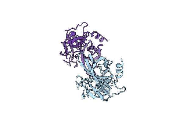
Organism: Mus musculus, Homo sapiens
Method: X-RAY DIFFRACTION Resolution:2.40 Å Release Date: 2007-12-11 Classification: PROTEIN BINDING Ligands: ZN |
 |
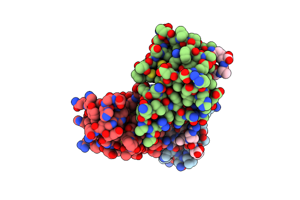
Organism: Mus musculus, Homo sapiens
Method: X-RAY DIFFRACTION Resolution:2.00 Å Release Date: 2007-12-11 Classification: PROTEIN BINDING Ligands: ZN |
 |
Organism: Mus musculus
Method: X-RAY DIFFRACTION Resolution:2.05 Å Release Date: 2007-12-11 Classification: PROTEIN BINDING Ligands: ZN |
 |
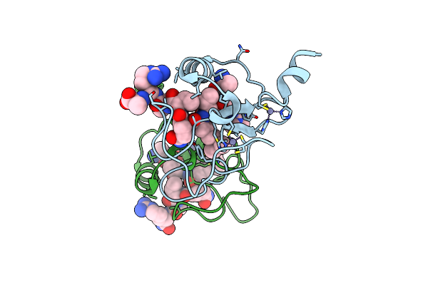
Organism: Mus musculus
Method: X-RAY DIFFRACTION Resolution:1.80 Å Release Date: 2007-12-11 Classification: PROTEIN BINDING Ligands: ZN |
 |
Organism: Mus musculus, Homo sapiens
Method: X-RAY DIFFRACTION Resolution:2.00 Å Release Date: 2007-12-11 Classification: PROTEIN BINDING Ligands: ZN |
 |
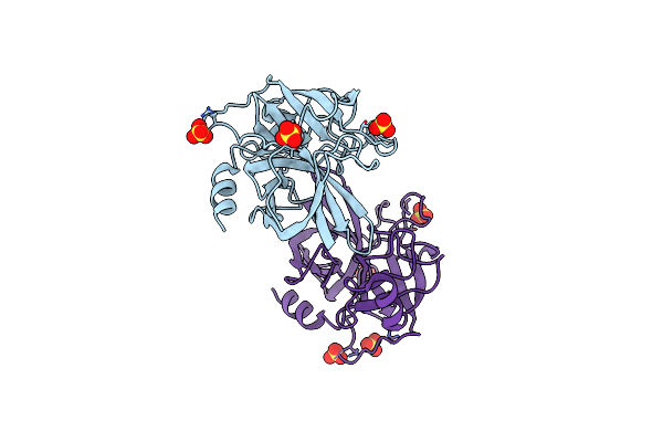
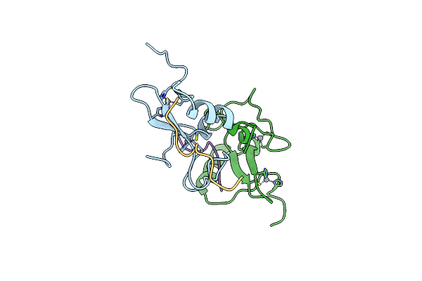
Crystal Structure Of Rag2-Phd Finger In Complex With H3K4Me3 Peptide At 1.1A Resolution
Organism: Mus musculus, Homo sapiens
Method: X-RAY DIFFRACTION Resolution:1.10 Å Release Date: 2007-12-11 Classification: PROTEIN BINDING Ligands: ZN |