
Search Count: 35
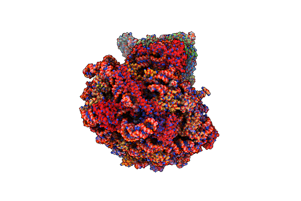 |
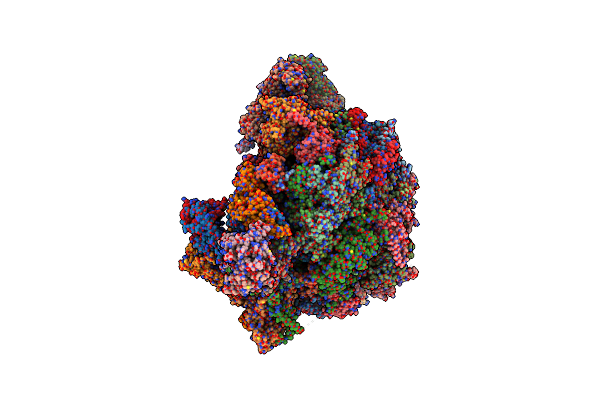
Organism: Schizosaccharomyces pombe
Method: ELECTRON MICROSCOPY Release Date: 2024-10-16 Classification: RIBOSOME Ligands: ZN |
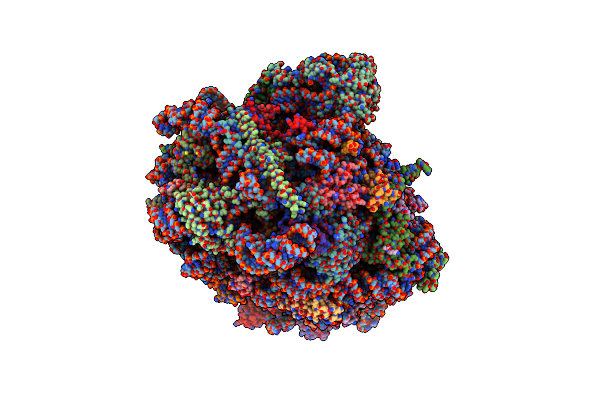 |
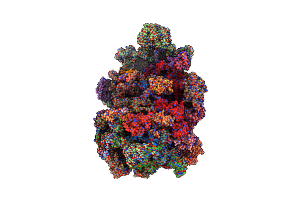
Organism: Schizosaccharomyces pombe
Method: ELECTRON MICROSCOPY Release Date: 2024-10-16 Classification: RIBOSOME Ligands: ZN, MG |
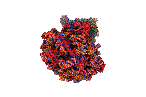 |
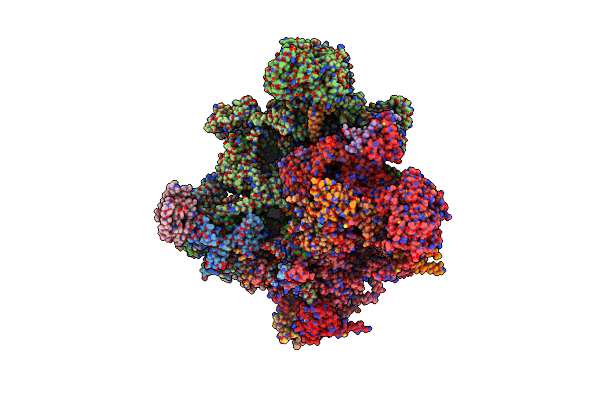
Organism: Schizosaccharomyces pombe
Method: ELECTRON MICROSCOPY Release Date: 2024-10-16 Classification: RIBOSOME Ligands: ZN |
 |
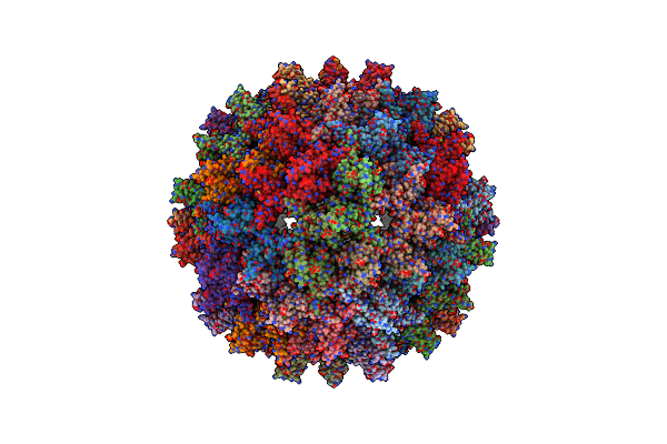
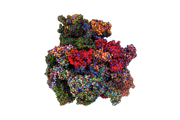
Organism: Mus musculus
Method: ELECTRON MICROSCOPY Release Date: 2023-09-20 Classification: HYDROLASE Ligands: PO4 |
 |
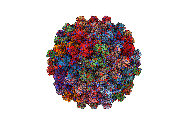
Organism: African cichlid nackednavirus
Method: ELECTRON MICROSCOPY Release Date: 2023-04-05 Classification: VIRAL PROTEIN |
 |
Organism: African cichlid nackednavirus
Method: ELECTRON MICROSCOPY Release Date: 2023-04-05 Classification: VIRAL PROTEIN |
 |
Organism: Escherichia coli b
Method: ELECTRON MICROSCOPY Release Date: 2022-11-30 Classification: RIBOSOME Ligands: ZN, K, MG |
 |
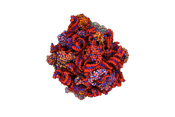
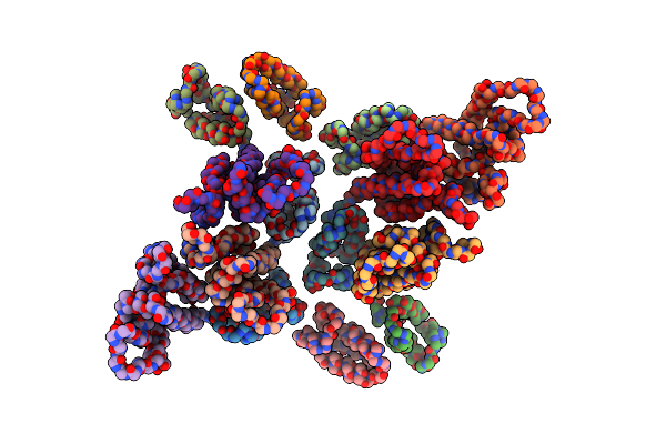
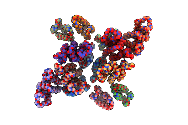
Structure Of Transcription Factor Uaf In Complex With Tbp And 35S Rrna Promoter Dna
Organism: Saccharomyces cerevisiae
Method: ELECTRON MICROSCOPY Release Date: 2022-04-27 Classification: TRANSCRIPTION |
 |
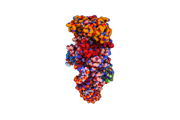
Mammalian Ribosome Nascent Chain Complex With Srp And Srp Receptor In Early State A
Organism: Homo sapiens, Oryctolagus cuniculus
Method: ELECTRON MICROSCOPY Release Date: 2021-06-02 Classification: RIBOSOME Ligands: ZN, MG, GTP, GNP |
 |
Organism: Apis mellifera
Method: ELECTRON MICROSCOPY Release Date: 2020-12-30 Classification: PROTEIN FIBRIL Ligands: 94R, NAG, SO4 |
 |
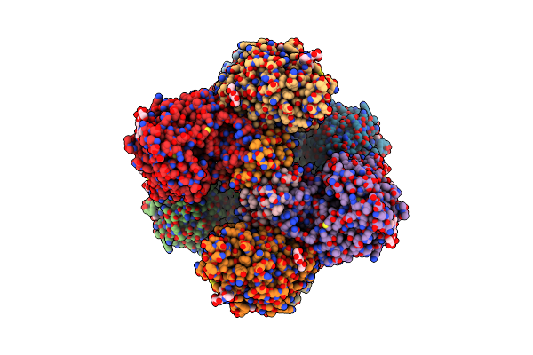
State A Of The Trypanosoma Brucei Mitoribosomal Large Subunit Assembly Intermediate
Organism: Trypanosoma brucei brucei
Method: ELECTRON MICROSCOPY Release Date: 2020-10-14 Classification: RIBOSOME Ligands: ZN, MG, GTP, NA, ATP, PM8, NAD |
 |
State B Of The Trypanosoma Brucei Mitoribosomal Large Subunit Assembly Intermediate
Organism: Trypanosoma brucei brucei
Method: ELECTRON MICROSCOPY Release Date: 2020-10-14 Classification: RIBOSOME Ligands: ZN, MG, SPD, GTP, NA, ATP, PM8, NAD |
 |
Organism: Trypanosoma brucei brucei
Method: ELECTRON MICROSCOPY Release Date: 2019-09-25 Classification: RIBOSOME Ligands: MG, PO4, PM8, ZN, GTP, SAH |
 |
Organism: Trypanosoma brucei brucei, Trypanosoma brucei brucei
Method: ELECTRON MICROSCOPY Release Date: 2019-09-18 Classification: RIBOSOME Ligands: GTP, MG, ZN, SAH |
 |
Organism: Trypanosoma brucei brucei
Method: ELECTRON MICROSCOPY Release Date: 2019-09-18 Classification: RIBOSOME Ligands: MG, PO4, PM8, ZN |
 |
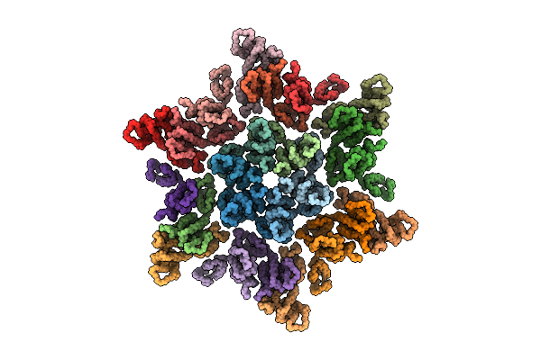
The Structure Of The Mature Hiv-1 Ca Hexameric Lattice With Curvature Parameters: Tilt=23, Twist=-6
Organism: Human immunodeficiency virus 1
Method: ELECTRON MICROSCOPY Release Date: 2017-01-18 Classification: VIRAL PROTEIN |
 |
Organism: Human immunodeficiency virus 1
Method: ELECTRON MICROSCOPY Release Date: 2016-12-28 Classification: VIRAL PROTEIN |
 |
Organism: Human immunodeficiency virus 1
Method: ELECTRON MICROSCOPY Release Date: 2016-12-28 Classification: VIRAL PROTEIN |
 |
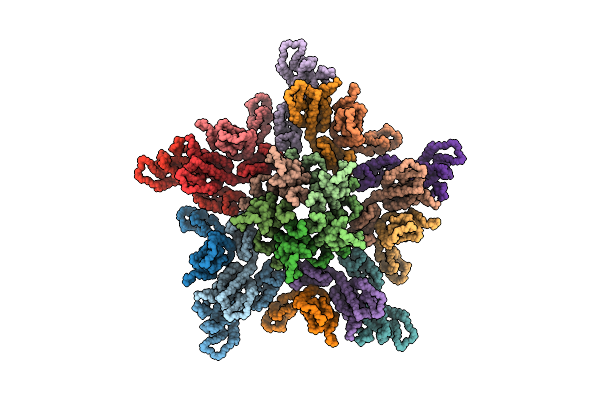
The Structure Of The Mature Hiv-1 Ca Hexameric Lattice With Curvature Parameters: Tilt=-1, Twist=0
Organism: Human immunodeficiency virus 1
Method: ELECTRON MICROSCOPY Release Date: 2016-12-28 Classification: VIRAL PROTEIN |
 |
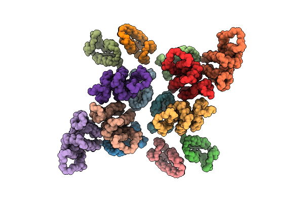
The Structure Of The Mature Hiv-1 Ca Hexameric Lattice With Curvature Parameters: Tilt=5, Twist=6
Organism: Human immunodeficiency virus 1
Method: ELECTRON MICROSCOPY Release Date: 2016-12-28 Classification: VIRAL PROTEIN |