Planned Maintenance: Some services may turn out to be unavailable from 15th January, 2026 to 16th January, 2026. We apologize for the inconvenience!

Planned Maintenance: Some services may turn out to be unavailable from 15th January, 2026 to 16th January, 2026. We apologize for the inconvenience!
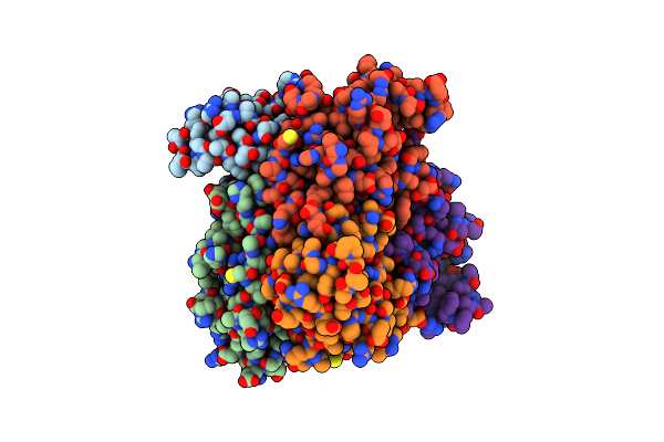 |
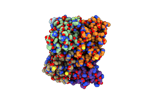
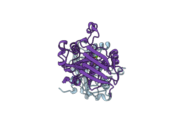
Organism: Homo sapiens
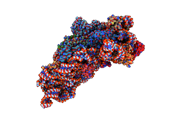
Method: X-RAY DIFFRACTION Resolution:2.64 Å Release Date: 2024-09-18 Classification: TRANSFERASE Ligands: PO4 |
 |
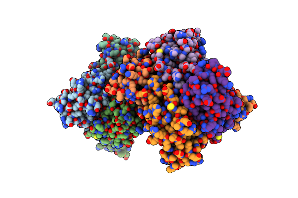
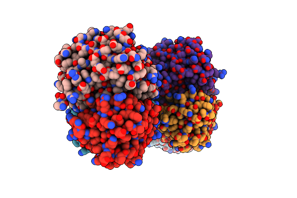
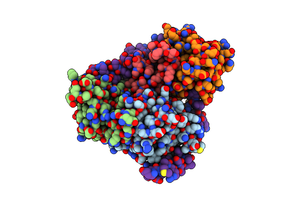
Organism: Homo sapiens
Method: X-RAY DIFFRACTION Resolution:1.77 Å Release Date: 2024-09-18 Classification: TRANSFERASE Ligands: CMP, SO4 |
 |
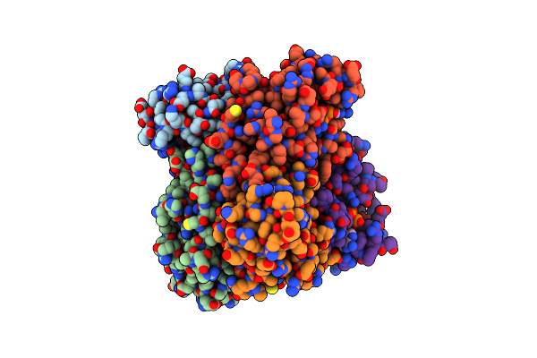
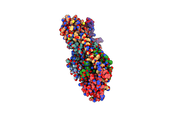
Organism: Homo sapiens
Method: X-RAY DIFFRACTION Resolution:2.18 Å Release Date: 2024-09-18 Classification: TRANSFERASE Ligands: GDP, SO4 |
 |
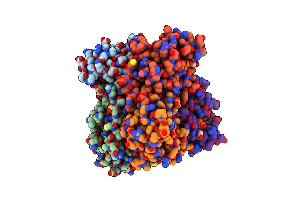
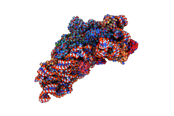
Organism: Homo sapiens
Method: X-RAY DIFFRACTION Resolution:2.10 Å Release Date: 2024-09-18 Classification: TRANSFERASE Ligands: ADP, MG |
 |
Organism: Homo sapiens
Method: X-RAY DIFFRACTION Resolution:1.87 Å Release Date: 2024-09-18 Classification: TRANSFERASE Ligands: UDP, MG |
 |
Organism: Homo sapiens
Method: X-RAY DIFFRACTION Resolution:1.26 Å Release Date: 2024-09-18 Classification: TRANSFERASE Ligands: ADP, SO4 |
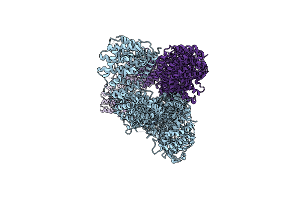 |
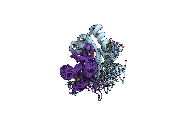
Organism: Homo sapiens
Method: ELECTRON MICROSCOPY Release Date: 2022-03-30 Classification: SIGNALING PROTEIN |
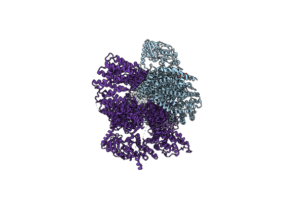 |
Organism: Homo sapiens
Method: ELECTRON MICROSCOPY Release Date: 2022-03-30 Classification: SIGNALING PROTEIN Ligands: GSP |
 |
X-Ray Structure Of Homo Sapiens Fumarylacetoacetate Hydrolase Domain Containing Protein 1 (Fahd1) In Complex With Inhibitor Oxalate At 1.94A Resolution.
Organism: Homo sapiens
Method: X-RAY DIFFRACTION Resolution:1.94 Å Release Date: 2018-11-07 Classification: hydrolase/lyase Ligands: MG, OXL, CL |
 |
X-Ray Structure Of Homo Sapiens Fumarylacetoacetate Hydrolase Domain Containing Protein 1 (Fahd1) At 1.56A Resolution.
Organism: Homo sapiens
Method: X-RAY DIFFRACTION Resolution:1.56 Å Release Date: 2018-11-07 Classification: HYDROLASE/LYASE Ligands: CL, MG |
 |
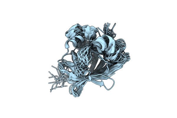
Organism: Homo sapiens
Method: X-RAY DIFFRACTION Resolution:2.30 Å Release Date: 2017-10-04 Classification: SIGNALING PROTEIN Ligands: CL |
 |
Organism: Homo sapiens
Method: X-RAY DIFFRACTION Resolution:2.90 Å Release Date: 2017-10-04 Classification: SIGNALING PROTEIN |
 |
Organism: Thermus thermophilus hb8
Method: X-RAY DIFFRACTION Resolution:2.90 Å Release Date: 2013-08-07 Classification: RIBOSOME Ligands: MG, K, ON0, ZN |
 |
Organism: Thermus thermophilus hb8
Method: X-RAY DIFFRACTION Resolution:3.00 Å Release Date: 2013-08-07 Classification: RIBOSOME Ligands: MG, K, M5Z, ZN |
 |
Organism: Thermus thermophilus hb8
Method: X-RAY DIFFRACTION Resolution:3.15 Å Release Date: 2013-08-07 Classification: RIBOSOME Ligands: MG, K, RPO, ZN |
 |
Organism: Thermus thermophilus hb8
Method: X-RAY DIFFRACTION Resolution:3.00 Å Release Date: 2013-08-07 Classification: RIBOSOME Ligands: MG, K, 3TS, ZN |
 |
Organism: Thermus thermophilus
Method: X-RAY DIFFRACTION Resolution:3.50 Å Release Date: 2012-07-18 Classification: RIBOSOME Ligands: MG, K, AM2, ZN |
 |
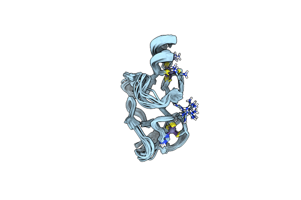
Organism: Homo sapiens
Method: SOLUTION NMR Release Date: 2006-11-21 Classification: LIGASE Ligands: ZN |
 |
Cell Transformation By The Myc Oncogene Activates Expression Of A Lipocalin: Analysis Of The Gene (Q83) And Solution Structure Of Its Protein Product
Organism: Coturnix coturnix
Method: SOLUTION NMR Release Date: 2003-07-15 Classification: LIPID BINDING PROTEIN |
 |
Quail Cysteine And Glycine-Rich Protein, Nmr, 15 Minimized Model Structures
Organism: Coturnix japonica
Method: SOLUTION NMR Release Date: 2001-09-05 Classification: METAL BINDING PROTEIN Ligands: ZN |