
Search Count: 33
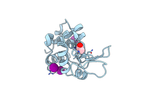 |
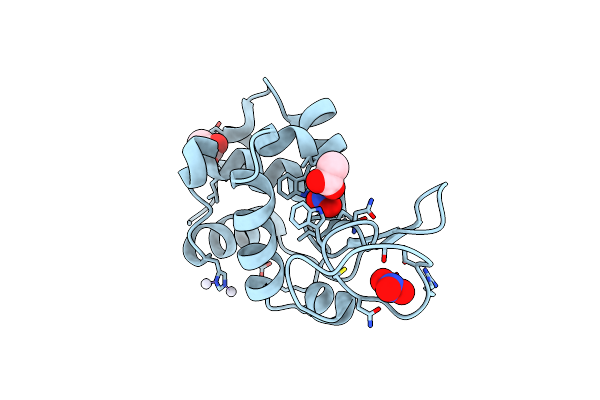
X-Ray Structure Of The Adduct Formed Upon Reaction Of The Diiodido Analogue Of Picoplatin With Lysozyme (Structure A)
Organism: Gallus gallus
Method: X-RAY DIFFRACTION Resolution:1.96 Å Release Date: 2025-05-14 Classification: HYDROLASE Ligands: NH3, ACT, CL, PT, IOD |
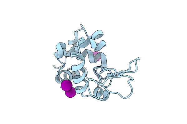 |
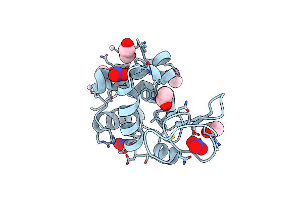
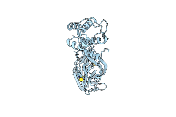
X-Ray Structure Of The Adduct Formed Upon Reaction Of The Diiodido Analogue Of Picoplatin With Lysozyme (Structure B)
Organism: Gallus gallus
Method: X-RAY DIFFRACTION Resolution:1.48 Å Release Date: 2025-05-14 Classification: HYDROLASE Ligands: PT, IOD |
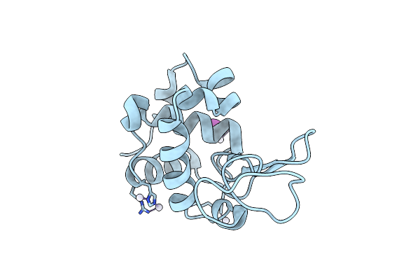 |
X-Structure Of The Adduct Formed Upon Reaction Of The Diiodido Analogue Of Picoplatin With Lysozyme (Structure C)
Organism: Gallus gallus
Method: X-RAY DIFFRACTION Resolution:2.25 Å Release Date: 2025-05-14 Classification: HYDROLASE Ligands: PT, NH3, IOD |
 |
X-Ray Structure Of The Adduct Formed Upon Reaction Of The Diiodido Analogue Of Picoplatin With Ribonuclease A
Organism: Bos taurus
Method: X-RAY DIFFRACTION Resolution:1.77 Å Release Date: 2025-05-14 Classification: HYDROLASE Ligands: NH3, PT, IOD, CL |
 |
X-Ray Structure Of The Adduct Formed Upon Reaction Of The Diiodido Analogue Of Picoplatin With Human Serum Albumin
Organism: Homo sapiens
Method: X-RAY DIFFRACTION Resolution:3.90 Å Release Date: 2025-05-14 Classification: HYDROLASE Ligands: PT |
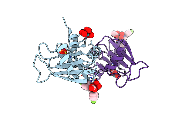 |
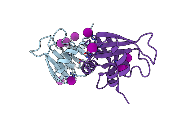
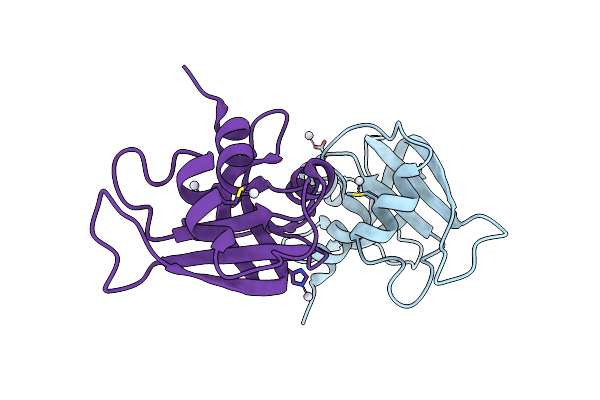
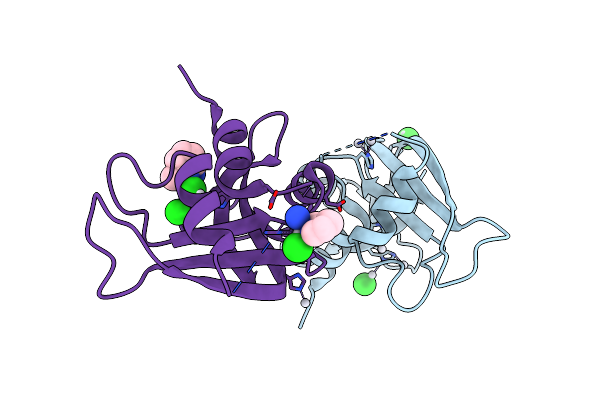
X-Ray Structure Of The Adduct Formed Upon Reaction Of Rnase A With [Ru2(D-P-Fphf)(O2Cch3)2(O2Co)] Complex
Organism: Bos taurus
Method: X-RAY DIFFRACTION Resolution:1.74 Å Release Date: 2024-12-11 Classification: STRUCTURAL PROTEIN Ligands: SO4, A1IQW |
 |
X-Ray Structure Of The Adduct Formed Upon Reaction Of Picoplatin With Lysozyme (Structure A)
Organism: Gallus gallus
Method: X-RAY DIFFRACTION Resolution:1.60 Å Release Date: 2024-05-22 Classification: HYDROLASE Ligands: NO3, ACT, NH3, PT |
 |
X-Ray Structure Of The Adduct Formed Upon Reaction Of Picoplatin With Lysozyme (Structure B)
Organism: Gallus gallus
Method: X-RAY DIFFRACTION Resolution:1.36 Å Release Date: 2024-05-22 Classification: HYDROLASE Ligands: NO3, GOL, ACT, PT |
 |
X-Ray Structure Of The Adduct Formed Upon Reaction Of Picoplatin With Bovine Pancreatic Ribonuclease (Structure C)
Organism: Gallus gallus
Method: X-RAY DIFFRACTION Resolution:1.99 Å Release Date: 2024-05-22 Classification: RNA BINDING PROTEIN Ligands: NH3, PT |
 |
X-Ray Structure Of The Adduct Formed Upon Reaction Of Picoplatin With Bovine Pancreatic Ribonuclease (Structure D)
Organism: Gallus gallus
Method: X-RAY DIFFRACTION Resolution:1.76 Å Release Date: 2024-05-22 Classification: RNA BINDING PROTEIN Ligands: NH3, PT, CL, A1H58 |
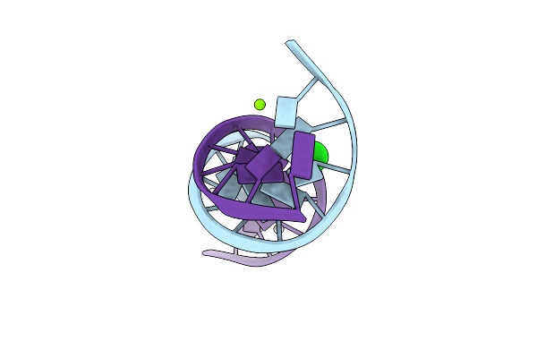 |
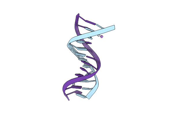
Crystal Structure Of Transplatin/B-Dna Adduct Obtained Upon 7 Days Of Soaking
Organism: Synthetic construct
Method: X-RAY DIFFRACTION Resolution:1.40 Å Release Date: 2024-02-28 Classification: DNA Ligands: MG, PT, NH3, CL |
 |
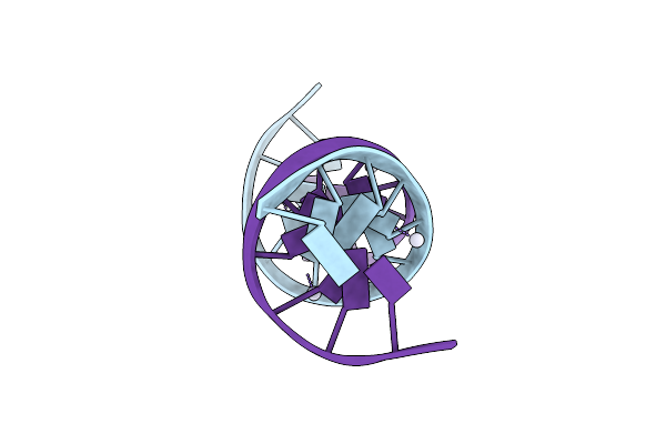
Crystal Structure Of Transplatin/B-Dna Adduct Obtained Upon 48 H Of Soaking
Organism: Dna molecule
Method: X-RAY DIFFRACTION Resolution:1.42 Å Release Date: 2024-02-07 Classification: DNA Ligands: MG, NH3, PT |
 |
Organism: Synthetic construct
Method: X-RAY DIFFRACTION Resolution:2.31 Å Release Date: 2024-01-31 Classification: DNA Ligands: PT |
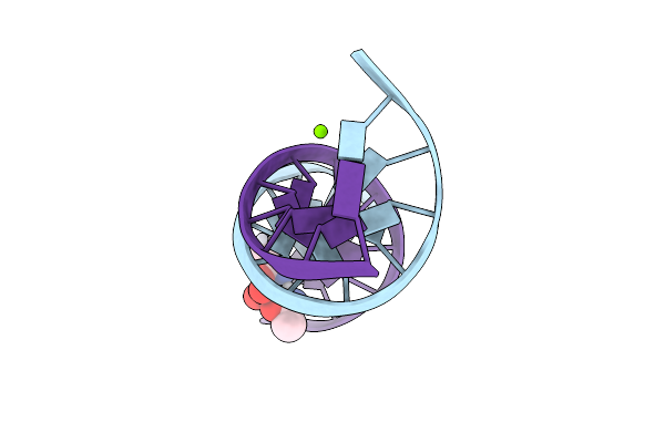 |
Crystal Structure Of Arsenoplatin-1/B-Dna Adduct Obtained Upon 4 H Of Soaking
Organism: Synthetic construct
Method: X-RAY DIFFRACTION Resolution:1.52 Å Release Date: 2024-01-31 Classification: DNA Ligands: PT, MG, A6R |
 |
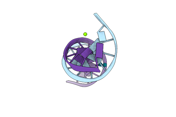
Crystal Structure Of Arsenoplatin-1/B-Dna Adduct Obtained Upon 48 H Of Soaking
Organism: Synthetic construct
Method: X-RAY DIFFRACTION Resolution:2.51 Å Release Date: 2024-01-31 Classification: DNA Ligands: PT, A6R |
 |
X-Ray Structure Of The Adduct Formed Upon Reaction Of A B-Dna Double Helical Dodecamer With Dirhodium Tetraacetate
Organism: Dna molecule
Method: X-RAY DIFFRACTION Resolution:1.24 Å Release Date: 2023-05-31 Classification: DNA Ligands: MG, RH, CL |
 |
Organism: Severe acute respiratory syndrome coronavirus 2
Method: X-RAY DIFFRACTION Resolution:2.42 Å Release Date: 2022-12-28 Classification: VIRAL PROTEIN Ligands: AU |
 |
Organism: Severe acute respiratory syndrome coronavirus 2
Method: X-RAY DIFFRACTION Resolution:2.40 Å Release Date: 2022-12-28 Classification: VIRAL PROTEIN Ligands: AU |
 |
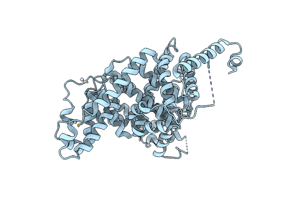
X-Ray Crystal Structure Of Sporosarcina Pasteurii Urease Inhibited By The Gold(I)-Phosphine Compound Au(Pet3)I Determined At 1.80 Angstroms
Organism: Sporosarcina pasteurii
Method: X-RAY DIFFRACTION Resolution:1.80 Å Release Date: 2022-06-01 Classification: HYDROLASE Ligands: EDO, SO4, NI, O, AUF, AU |
 |
X-Ray Crystal Structure Of Sporosarcina Pasteurii Urease Inhibited By The Gold(I)-Diphosphine Compound Au(Pet3)2Cl Determined At 1.87 Angstroms
Organism: Sporosarcina pasteurii
Method: X-RAY DIFFRACTION Resolution:1.87 Å Release Date: 2022-06-01 Classification: HYDROLASE Ligands: EDO, SO4, NI, O, AUF |