
Search Count: 140
 |
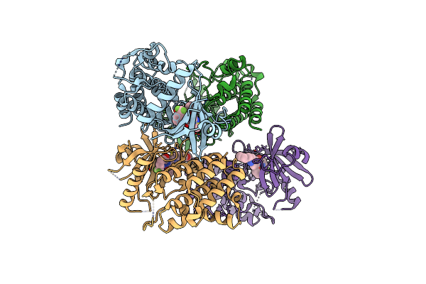
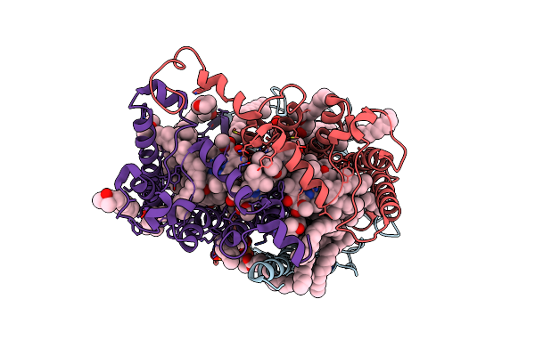
Glucagon Like Peptide Receptor-1 (Glp1R) A316T Mutant With Glp-1 Peptide. Dominant Negative Gs Complex.
Organism: Homo sapiens, Lama glama
Method: ELECTRON MICROSCOPY Release Date: 2025-10-29 Classification: MEMBRANE PROTEIN |
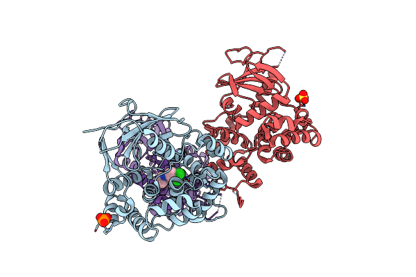 |
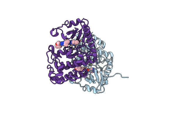
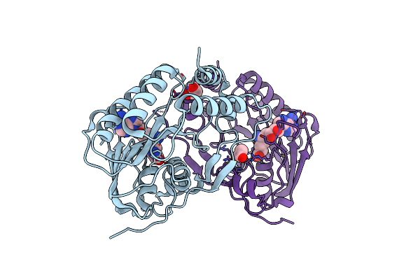
Structure Of The Mouse 8-Oxoguanine Dna Glycosylase Mogg1 In Complex With Ligand Th14445
Organism: Mus musculus
Method: X-RAY DIFFRACTION Release Date: 2025-05-21 Classification: DNA BINDING PROTEIN Ligands: A1IVU, PO4, NI, SO4 |
 |
Organism: Solanum tuberosum
Method: X-RAY DIFFRACTION Resolution:1.65 Å Release Date: 2025-05-14 Classification: PLANT PROTEIN Ligands: GOL, ETE, MAN |
 |
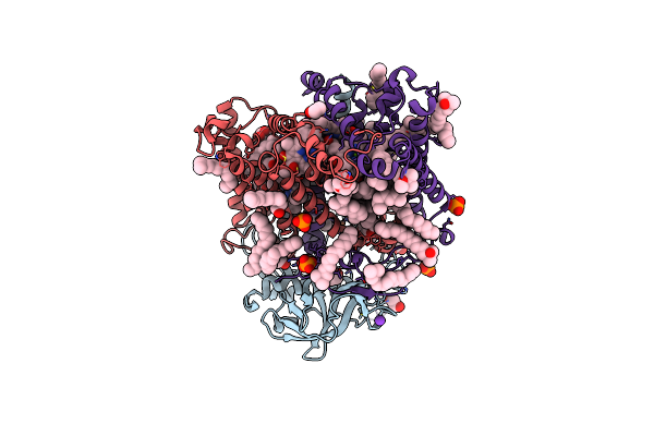
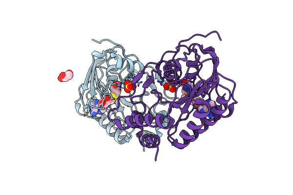
Complex Between The Porcine Trypsin And M271 A Kunitz-Sti From Solanum Tuberosum
Organism: Solanum tuberosum, Sus scrofa
Method: X-RAY DIFFRACTION Resolution:2.42 Å Release Date: 2025-05-14 Classification: HYDROLASE Ligands: CA |
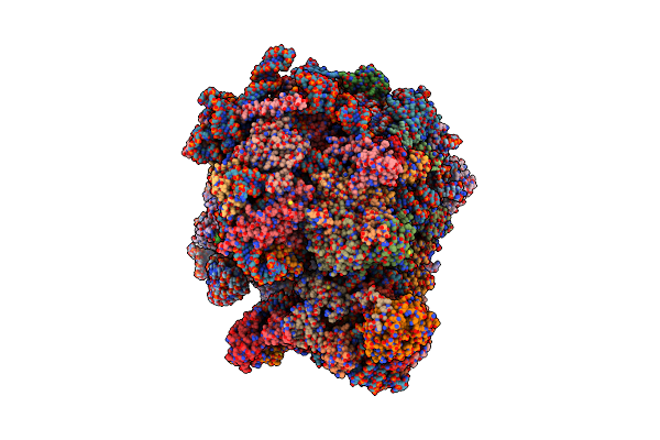 |
Structure Of Candida Albicans 80S Ribosome In Complex With Mefloquine (Non-Rotated State)
Organism: Candida albicans sc5314
Method: ELECTRON MICROSCOPY Release Date: 2025-04-23 Classification: RIBOSOME Ligands: ZN, YMZ, SPK |
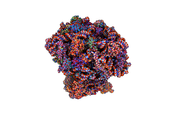 |
The Structure Of The Candida Albicans Ribosome With Trna-Fmet, Mrna, And Compounds (Gen And Mfq) Shows Strong Density For The A Site Trna
Organism: Candida albicans
Method: ELECTRON MICROSCOPY Release Date: 2025-04-23 Classification: RIBOSOME Ligands: SPK, GET, ZN |
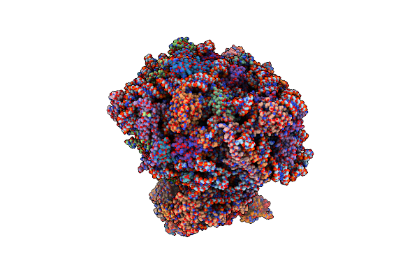 |
The Structure Of The Candida Albicans Ribosome With Trna-Fmet, Mrna, And Compounds (Gen And Mfq) With Strong Density For The P-Site Trna
Organism: Escherichia coli, Candida albicans sc5314
Method: ELECTRON MICROSCOPY Release Date: 2025-04-23 Classification: RIBOSOME Ligands: SPK, GET, ZN, YMZ |
 |
Organism: Homo sapiens
Method: X-RAY DIFFRACTION Resolution:1.85 Å Release Date: 2024-06-26 Classification: TRANSFERASE/INHIBITOR Ligands: X1T |
 |
Organism: Homo sapiens
Method: X-RAY DIFFRACTION Resolution:1.80 Å Release Date: 2024-06-26 Classification: TRANSFERASE/INHIBITOR Ligands: WFQ |
 |
Double Mutant A(L37)C/S(L99)C Structure Of Photosynthetic Reaction Center From Cereibacter Sphaeroides Strain Rv
Organism: Cereibacter sphaeroides 2.4.1
Method: X-RAY DIFFRACTION Resolution:2.60 Å Release Date: 2023-11-22 Classification: PHOTOSYNTHESIS Ligands: LDA, UNL, PO4, EDO, K, BCL, BPH, U10, DIO, HTO, CDL, FE, SPN |
 |
Double Mutant A(L53)C/I(L64)C Structure Of Photosynthetic Reaction Center From Cereibacter Sphaeroides Strain Rv
Organism: Cereibacter sphaeroides 2.4.1
Method: X-RAY DIFFRACTION Resolution:2.86 Å Release Date: 2023-11-22 Classification: PHOTOSYNTHESIS Ligands: LDA, UNL, PO4, EDO, K, BCL, BPH, U10, DIO, HTO, CDL, FE, SPN |
 |
Double Mutant V(M84)C/A(L278)C Structure Of Photosynthetic Reaction Center From Cereibacter Sphaeroides Strain Rv
Organism: Cereibacter sphaeroides 2.4.1
Method: X-RAY DIFFRACTION Resolution:2.60 Å Release Date: 2023-11-22 Classification: PHOTOSYNTHESIS Ligands: LDA, UNL, EDO, BCL, BPH, HTO, CDL, FE, U10, SPN |
 |
Double Mutant A(L172)C/L(L246)C Structure Of Photosynthetic Reaction Center From Cereibacter Sphaeroides Strain Rv
Organism: Cereibacter sphaeroides 2.4.1
Method: X-RAY DIFFRACTION Resolution:2.45 Å Release Date: 2023-11-22 Classification: PHOTOSYNTHESIS Ligands: LDA, UNL, EDO, OLC, BCL, BPH, HTO, CDL, FE, U10, SPN |
 |
Double Mutant G(M19)C/T(L214)C Structure Of Photosynthetic Reaction Center From Cereibacter Sphaeroides Strain Rv
Organism: Cereibacter sphaeroides 2.4.1
Method: X-RAY DIFFRACTION Resolution:2.75 Å Release Date: 2023-11-22 Classification: PHOTOSYNTHESIS Ligands: NKP, LDA, UNL, OLC, BCL, BPH, DIO, EDO, FE, U10, SPN, PO4 |
 |
Organism: Homo sapiens
Method: X-RAY DIFFRACTION Release Date: 2023-11-22 Classification: TRANSFERASE Ligands: XRU, SO4, EDO, UNX |
 |
Organism: Usutu virus
Method: X-RAY DIFFRACTION Resolution:1.84 Å Release Date: 2023-10-04 Classification: VIRAL PROTEIN Ligands: SFG, GOL, GLY |
 |
Organism: Usutu virus
Method: X-RAY DIFFRACTION Resolution:2.22 Å Release Date: 2023-10-04 Classification: VIRAL PROTEIN Ligands: SO4, SAH, GOL, SAM |
 |
Organism: Synthetic construct, Candida albicans
Method: ELECTRON MICROSCOPY Release Date: 2023-09-13 Classification: RIBOSOME Ligands: SPK, K16, SPD, MG, ZN |
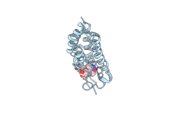 |
Crystal Structure Of The First Bromodomain Of Human Brd4 L94C Variant In Complex With Racemic 3,5-Dimethylisoxazol Ligand
Organism: Homo sapiens
Method: X-RAY DIFFRACTION Resolution:1.88 Å Release Date: 2023-08-02 Classification: GENE REGULATION Ligands: E5Q, V0R |
 |
Organism: Homo sapiens
Method: POWDER DIFFRACTION Resolution:2.29 Å Release Date: 2023-06-21 Classification: HORMONE |