
Search Count: 25
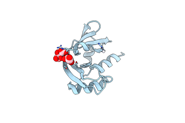 |
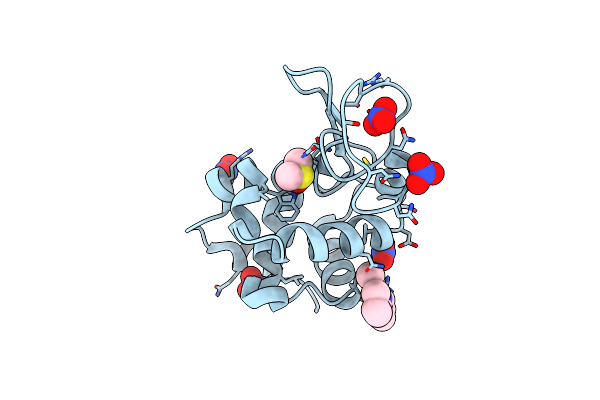
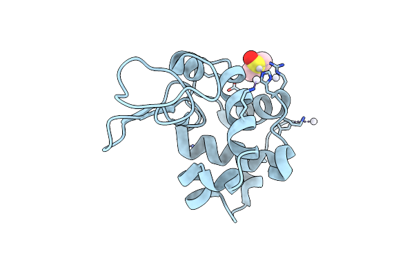
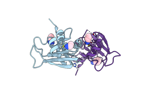
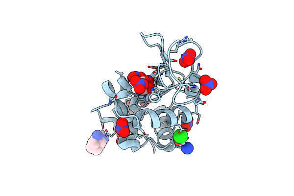
X-Ray Structure Of The Adduct Formed Upon Reaction Of Cisplatin With Human Angiogenin After 5 Days Soaking
Organism: Homo sapiens
Method: X-RAY DIFFRACTION Resolution:1.76 Å Release Date: 2023-07-12 Classification: HYDROLASE Ligands: GOL, NH3, PT, TAR |
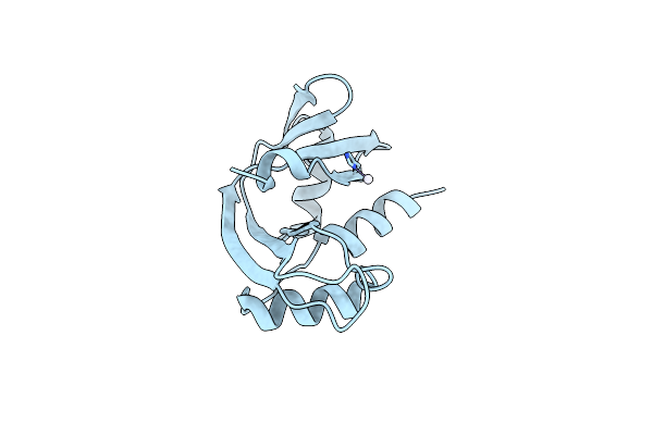 |
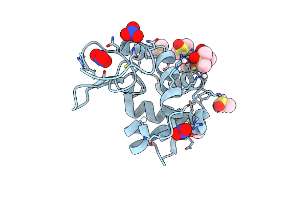
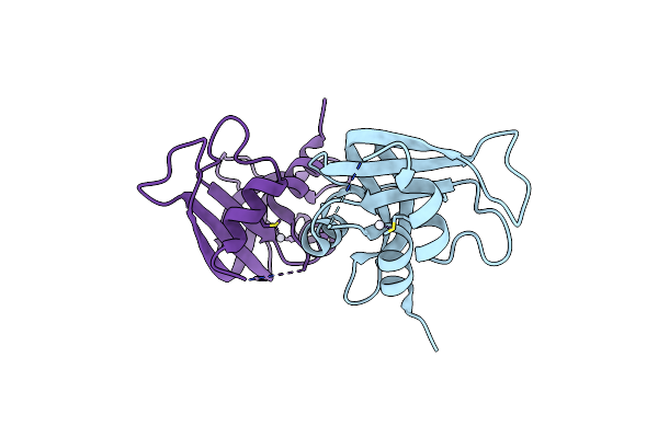
X-Ray Structure Of The Adduct Formed Upon Reaction Of Cisplatin With Human Angiogenin After 1 Month Soaking
Organism: Homo sapiens
Method: X-RAY DIFFRACTION Resolution:1.99 Å Release Date: 2023-07-12 Classification: HYDROLASE Ligands: NH3, PT |
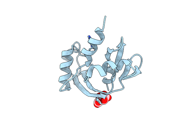 |
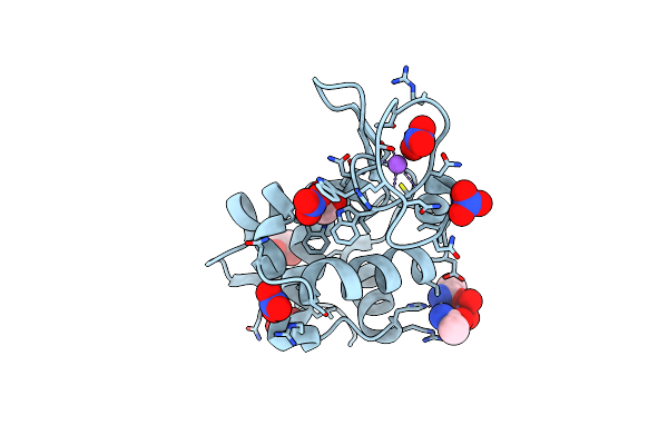
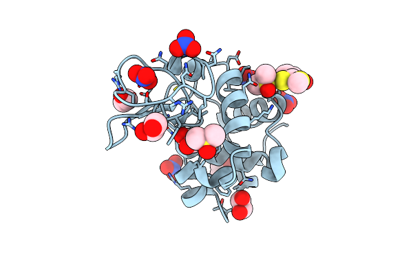
X-Ray Structure Of The Adduct Formed Upon Reaction Of Oxaliplatin With Human Angiogenin
Organism: Homo sapiens
Method: X-RAY DIFFRACTION Resolution:1.86 Å Release Date: 2022-03-23 Classification: HYDROLASE Ligands: PT, TAR |
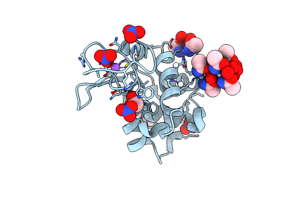 |
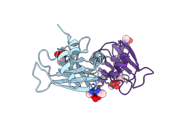
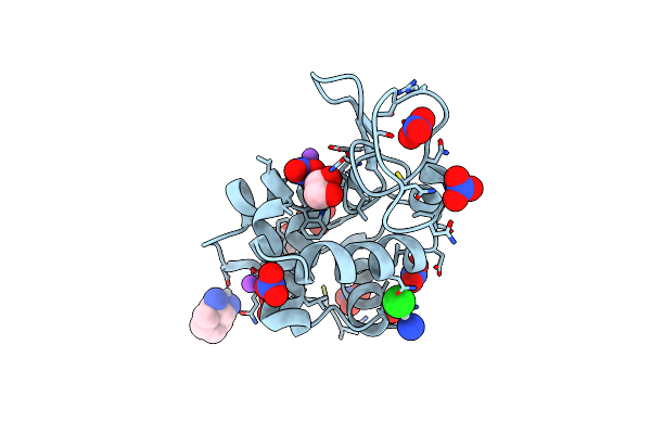
X-Ray Structure Of The Adduct Formed Upon Treating Lysozyme With An Aged Solution Of Arsenoplatin-1
Organism: Gallus gallus
Method: X-RAY DIFFRACTION Resolution:2.15 Å Release Date: 2020-12-23 Classification: HYDROLASE Ligands: NO3, EDO, A6R, DMS, NA |
 |
The Cytotoxic [Pt(H2Bapbpy)] Platinum Complex Interacting With The Cgtacg Hexamer
Organism: Synthetic construct
Method: X-RAY DIFFRACTION Resolution:2.30 Å Release Date: 2019-05-29 Classification: DNA Ligands: MG, PT9 |
 |
The X-Ray Structure Of The Adduct Formed In The Reaction Between Hen Egg White Lysozyme And Complex I, A Pentacoordinate Pt(Ii) Compound Containing 2,9-Dimethyl-1,10-Phenanthroline, Dimethylfumarate, Methyl And Iodine As Ligands
Organism: Gallus gallus
Method: X-RAY DIFFRACTION Resolution:1.96 Å Release Date: 2019-03-27 Classification: LYASE Ligands: NO3, J9H, DMS |
 |
The X-Ray Structure Of The Adduct Formed In The Reaction Between Bovine Pancreatic Ribonuclease And Complex I, A Pentacoordinate Pt(Ii) Compound Containing 2,9-Dimethyl-1,10-Phenanthroline, Dimethylfumarate, Methyl And Iodine As Ligands
Organism: Bos taurus
Method: X-RAY DIFFRACTION Resolution:2.03 Å Release Date: 2019-02-27 Classification: HYDROLASE Ligands: SO4, J9H |
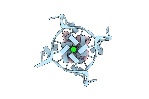 |
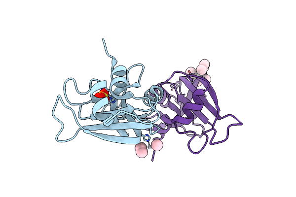
Structure Of The Complex Of A Human Telomeric Dna With Bis(1-Butyl-3-Methyl-Imidazole-2-Ylidene) Gold(I)
Organism: Homo sapiens
Method: X-RAY DIFFRACTION Resolution:2.00 Å Release Date: 2019-01-30 Classification: DNA Ligands: K, SR, FTQ |
 |
X-Ray Structure Of The Adduct Formed Upon Reaction Of Ribonuclease A With A Tetranuclear Pt-Thiosemicarbazone Compound
Organism: Bos taurus
Method: X-RAY DIFFRACTION Resolution:1.78 Å Release Date: 2018-08-29 Classification: HYDROLASE Ligands: PT, DMS |
 |
X-Ray Structure Of The Adduct Formed Upon Reaction Of Hen Egg White Lysozyme With A Tetranuclear Pt-Thiosemicarbazone Compound
Organism: Gallus gallus
Method: X-RAY DIFFRACTION Resolution:1.78 Å Release Date: 2018-08-29 Classification: HYDROLASE Ligands: EDO, NO3, DMS, PT |
 |
The X-Ray Structure Of The Adduct Formed In The Reaction Between Lysozyme And A Platinum(Ii) Terpyridine Compound (Ph 7.0)
Organism: Gallus gallus
Method: X-RAY DIFFRACTION Resolution:1.96 Å Release Date: 2018-07-04 Classification: HYDROLASE Ligands: PT, DMS, CL |
 |
The X-Ray Structure Of The Adduct Formed In The Reaction Between Lysozyme And A Platinum(Ii) Terpyridine Compound (Acid Ph)
Organism: Gallus gallus
Method: X-RAY DIFFRACTION Resolution:1.49 Å Release Date: 2018-06-27 Classification: HYDROLASE Ligands: NO3, PT, DMS |
 |
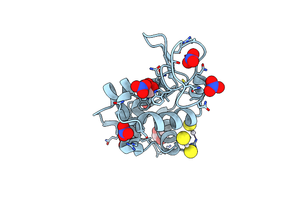
The X-Ray Structure Of The Adduct Formed In The Reaction Between Hen Egg White Lysozyme And Arsenoplatin-1
Organism: Gallus gallus
Method: X-RAY DIFFRACTION Resolution:1.85 Å Release Date: 2018-05-16 Classification: HYDROLASE Ligands: EDO, NO3, A6R, NA |
 |
The X-Ray Structure Of The Adduct Formed In The Reaction Between Bovine Pancreatic Ribonuclease And Arsenoplatin-1
Organism: Bos taurus
Method: X-RAY DIFFRACTION Resolution:2.15 Å Release Date: 2018-05-16 Classification: HYDROLASE Ligands: A6R |
 |
The X-Ray Structure Of The Adduct Formed In The Reaction Between Bovine Pancreatic Ribonuclease And Oxaliplatin
Organism: Bos taurus
Method: X-RAY DIFFRACTION Resolution:2.27 Å Release Date: 2015-11-25 Classification: HYDROLASE Ligands: 1PT |
 |
The X-Ray Structure Of The Adduct Formed In The Reaction Between Bovine Pancreatic Ribonuclease And Carboplatin
Organism: Bos taurus
Method: X-RAY DIFFRACTION Resolution:2.09 Å Release Date: 2015-11-18 Classification: HYDROLASE/RNA Ligands: QPT |
 |
X-Ray Structure Of The Adduct Formed In The Reaction Between Lysozyme And A Platinum(Ii) Complex With S,O Bidentate Ligands (9B)
Organism: Gallus gallus
Method: X-RAY DIFFRACTION Resolution:2.15 Å Release Date: 2015-09-02 Classification: HYDROLASE Ligands: 4KV, DMS, NO3, EDO |
 |
X-Ray Structure Of The Adduct Formed In The Reaction Between Lysozyme And A Platinum(Ii) Compound With A S,O Bidentate Ligand (9A=Chloro-(1-(3'-Hydroxy)-3-(Methylthio)-3-Thioxo-Prop-1-En-1-Olate-O,S)-(Dimethylsulfoxide-S)-Platinum(Ii))
Organism: Gallus gallus
Method: X-RAY DIFFRACTION Resolution:1.89 Å Release Date: 2015-09-02 Classification: HYDROLASE Ligands: 4KV, DMS, NO3, EDO |
 |
X-Ray Structure Of The Bis-Platinum Lysozyme Adduct Formed In The Reaction Between The Protein And The Two Drugs Cisplatin And Oxaliplatin
Organism: Gallus gallus
Method: X-RAY DIFFRACTION Resolution:1.85 Å Release Date: 2015-05-27 Classification: HYDROLASE Ligands: EDO, NO3, NA, 1PT, CPT |
 |
X-Ray Structure Of The Bis-Platinum Lysozyme Adduct Formed In The Reaction Between The Protein And The Two Drugs Cisplatin And Oxaliplatin (Preparation 2)
Organism: Gallus gallus
Method: X-RAY DIFFRACTION Resolution:1.95 Å Release Date: 2015-05-27 Classification: HYDROLASE Ligands: NO3, EDO, 1PT, CPT |