
Search Count: 15
 |
Organism: Vibrio cholerae
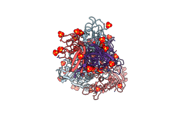
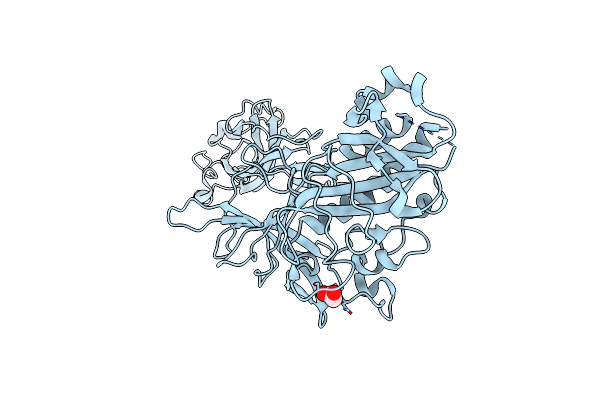
Method: X-RAY DIFFRACTION Resolution:2.32 Å Release Date: 2022-11-09 Classification: CELL ADHESION Ligands: SO4 |
 |
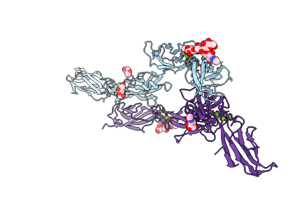
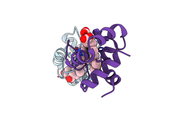
Crystal Structure Of Minor Pilin Tcpb From Vibrio Cholerae Complexed With N-Terminal Peptide Fragment Of Tcpf
Organism: Vibrio cholerae
Method: X-RAY DIFFRACTION Resolution:2.30 Å Release Date: 2022-11-09 Classification: CELL ADHESION Ligands: CA, CL, 1PE |
 |
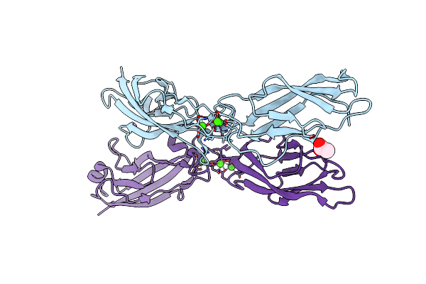
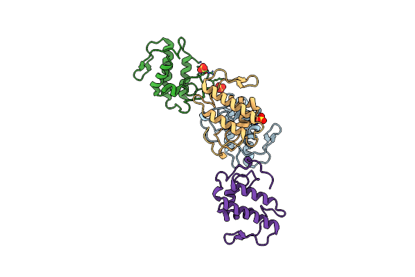
Crystal Structure Of Minor Pilin Tcpb From Vibrio Cholerae Complexed With Secreted Protein Tcpf
Organism: Vibrio cholerae
Method: X-RAY DIFFRACTION Resolution:4.05 Å Release Date: 2022-11-09 Classification: CELL ADHESION |
 |
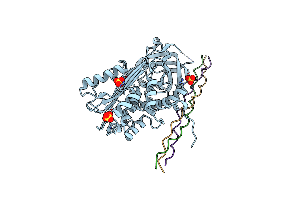
Organism: Homo sapiens
Method: X-RAY DIFFRACTION Resolution:2.70 Å Release Date: 2021-08-18 Classification: CELL ADHESION Ligands: NAG, CA |
 |
Organism: Homo sapiens
Method: X-RAY DIFFRACTION Resolution:1.38 Å Release Date: 2021-06-16 Classification: CELL ADHESION Ligands: CA, ACT, NA |
 |
Crystal Structure Of Mouse Pedf In Complex With Heterotrimeric Collagen Model Peptide.
Organism: Mus musculus, Homo sapiens
Method: X-RAY DIFFRACTION Resolution:2.48 Å Release Date: 2020-09-02 Classification: SIGNALING PROTEIN Ligands: SO4 |
 |
Organism: Shewanella benthica db6705
Method: X-RAY DIFFRACTION Resolution:1.71 Å Release Date: 2019-06-12 Classification: ELECTRON TRANSPORT Ligands: HEC, 1PE |
 |
Organism: Shewanella violacea (strain jcm 10179 / cip 106290 / lmg 19151 / dss12)
Method: X-RAY DIFFRACTION Resolution:2.14 Å Release Date: 2019-06-12 Classification: ELECTRON TRANSPORT Ligands: HEC |
 |
Organism: Protobothrops flavoviridis
Method: X-RAY DIFFRACTION Resolution:2.57 Å Release Date: 2019-01-16 Classification: TOXIN Ligands: SO4 |
 |
Crystal Structure Of Minor Pilin Cofb From Cfa/Iii Complexed With N-Terminal Peptide Fragment Of Cofj
Organism: Escherichia coli
Method: X-RAY DIFFRACTION Resolution:3.52 Å Release Date: 2018-06-27 Classification: CELL ADHESION |
 |
Organism: Escherichia coli
Method: X-RAY DIFFRACTION Resolution:1.76 Å Release Date: 2018-06-27 Classification: CELL ADHESION Ligands: CA |
 |
Homo-Dimeric Structure Of Cytochrome C' From Thermophilic Hydrogenophilus Thermoluteolus
Organism: Hydrogenophilus thermoluteolus
Method: X-RAY DIFFRACTION Resolution:1.89 Å Release Date: 2017-03-01 Classification: ELECTRON TRANSPORT Ligands: HEC |
 |
Organism: Shewanella violacea
Method: X-RAY DIFFRACTION Resolution:1.78 Å Release Date: 2016-10-19 Classification: ELECTRON TRANSPORT Ligands: HEC, IMD |
 |
The Crystal Structure Of Cofb, The Minor Pilin Subunit Of Cfa/Iii From Human Enterotoxigenic Escherichia Coli.
Organism: Escherichia coli
Method: X-RAY DIFFRACTION Resolution:1.88 Å Release Date: 2016-03-09 Classification: CELL ADHESION Ligands: ACT |
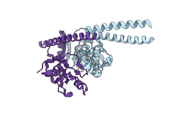 |
Crystal Structure Of The Human Condensin Smc Hinge Domain Heterodimer With Short Coiled Coils
Organism: Homo sapiens
Method: X-RAY DIFFRACTION Resolution:1.89 Å Release Date: 2015-08-26 Classification: PROTEIN BINDING |