
Search Count: 49
 |
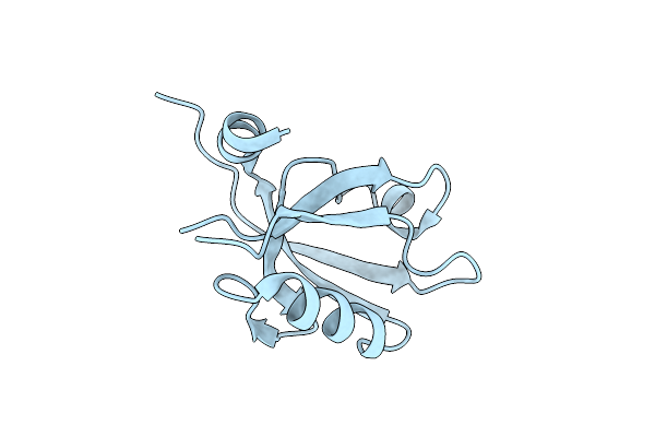
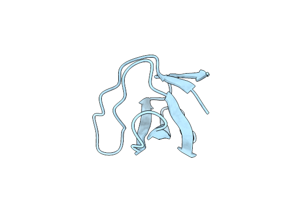
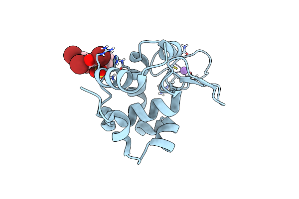
Crystal Structure Of The Third Pdz Domain Of Psd-95 Protein In The Space Group P3112 At Ph 4.0
Organism: Homo sapiens
Method: X-RAY DIFFRACTION Resolution:1.48 Å Release Date: 2023-03-08 Classification: PROTEIN BINDING Ligands: ACT |
 |
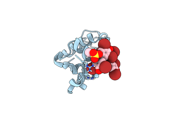
Crystal Structure Of The Third Pdz Domain Of Psd-95 Protein In The Space Group P212121 At Ph 4.6
Organism: Homo sapiens
Method: X-RAY DIFFRACTION Resolution:1.25 Å Release Date: 2023-03-08 Classification: PROTEIN BINDING Ligands: ACT |
 |
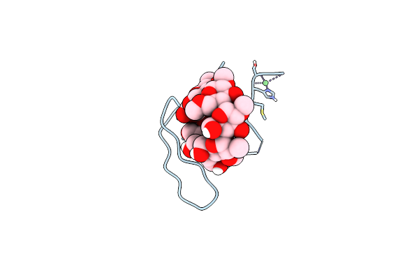
Crystal Structure Of The Third Pdz Domain Of Psd-95 Protein In The Space Group P21 At Ph 4.0
Organism: Homo sapiens
Method: X-RAY DIFFRACTION Resolution:1.63 Å Release Date: 2023-03-08 Classification: PROTEIN BINDING Ligands: ACT |
 |
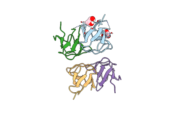
Crystal Structure Of The Third Pdz Domain Of Psd-95 Protein In The Space Group P212121 At Ph 4.0
Organism: Homo sapiens
Method: X-RAY DIFFRACTION Resolution:1.25 Å Release Date: 2023-03-08 Classification: PROTEIN BINDING Ligands: SO4 |
 |
Crystal Structure Of The Third Pdz Domain Of Psd-95 Protein In The Space Group P3121 At Ph 3.7
Organism: Homo sapiens
Method: X-RAY DIFFRACTION Resolution:1.50 Å Release Date: 2023-03-08 Classification: PROTEIN BINDING |
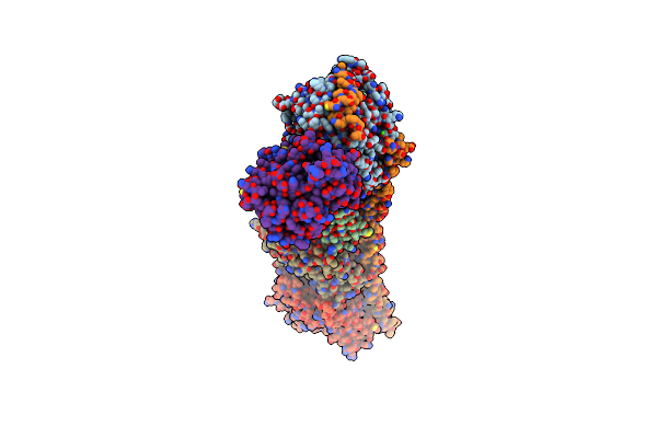 |
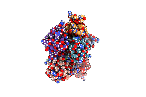
Organism: Rattus norvegicus, Gallus gallus, Bos taurus
Method: X-RAY DIFFRACTION Resolution:2.00 Å Release Date: 2022-12-14 Classification: CELL CYCLE Ligands: GTP, MG, CA, GOL, GDP, MES, KLC, ACP |
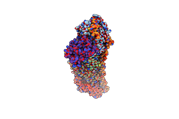 |
Organism: Rattus norvegicus, Gallus gallus, Bos taurus
Method: X-RAY DIFFRACTION Resolution:2.50 Å Release Date: 2022-11-23 Classification: CELL CYCLE/INHIBITOR Ligands: GTP, MG, GOL, CL, CA, GDP, MES, K9I, DMS, ACP |
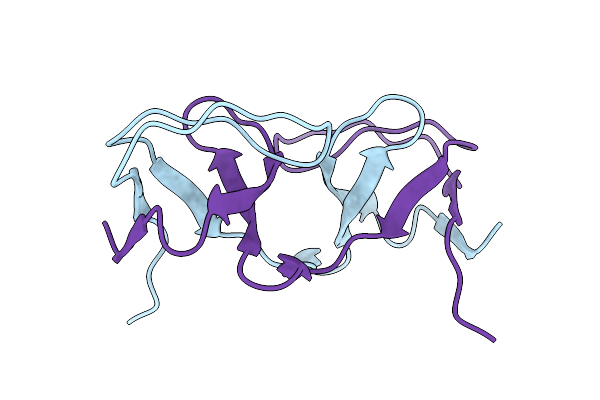 |
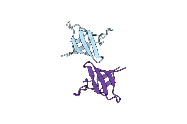
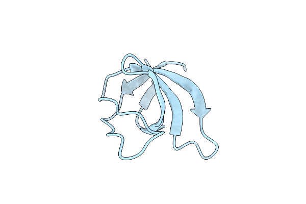
Crystal Structure Of The Intertwined Dimer Of The C-Src Sh3 Domain E93V-S94A-R95S-T96G Mutant
Organism: Gallus gallus
Method: X-RAY DIFFRACTION Release Date: 2022-11-16 Classification: PROTEIN BINDING |
 |
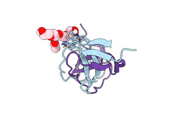
Crystal Structure Of The Abl Sh3 Domain V73E-A74S-S75R-G76T-D77E-G92N-Y93N-N94T-H95E Mutant In The Space Group P21221
Organism: Homo sapiens
Method: X-RAY DIFFRACTION Release Date: 2022-09-14 Classification: PROTEIN BINDING |
 |
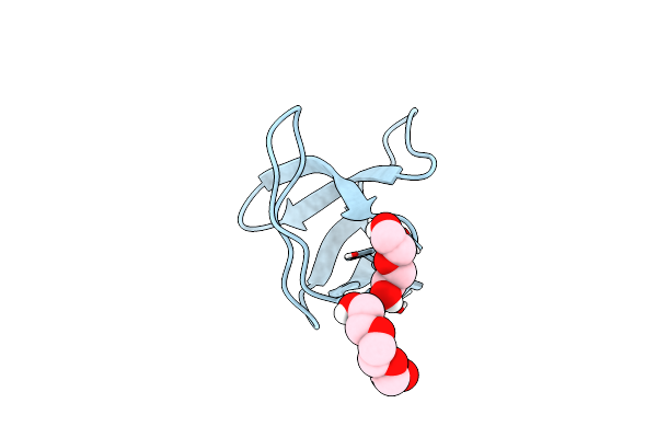
Crystal Structure Of The Abl Sh3 Domain V73E-A74S-S75R-G76T-D77E-G92N-Y93N-N94T-H95E Mutant In Presence Of Peg 200
Organism: Homo sapiens
Method: X-RAY DIFFRACTION Release Date: 2022-09-14 Classification: PROTEIN BINDING Ligands: PGE, NA, SO4, PEG, P6G |
 |
Organism: Homo sapiens
Method: X-RAY DIFFRACTION Release Date: 2022-09-14 Classification: PROTEIN BINDING Ligands: PEG, PGE |
 |
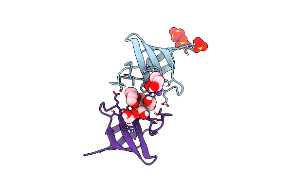
Crystal Structure Of The Intertwined Dimer Of The C-Src Sh3 Domain E93V-S94A-R95S-T96G-N112G-N113Y-T114N-E115H Mutant
Organism: Gallus gallus
Method: X-RAY DIFFRACTION Release Date: 2022-09-14 Classification: PROTEIN BINDING Ligands: PG4, ACT |
 |
Organism: Gallus gallus
Method: X-RAY DIFFRACTION Release Date: 2022-09-14 Classification: PROTEIN BINDING |
 |
Organism: Gallus gallus
Method: X-RAY DIFFRACTION Release Date: 2022-09-14 Classification: PROTEIN BINDING |
 |
Organism: Homo sapiens
Method: X-RAY DIFFRACTION Release Date: 2022-09-14 Classification: PROTEIN BINDING |
 |
Crystal Structure Of The Lysozyme In Presence Of Bromophenol Blue At Ph 6.5
Organism: Gallus gallus
Method: X-RAY DIFFRACTION Resolution:1.38 Å Release Date: 2020-09-09 Classification: HYDROLASE |
 |
Crystal Structure Of The Lysozyme In Presence Of Bromophenol Blue At Ph 5.5
Organism: Gallus gallus
Method: X-RAY DIFFRACTION Resolution:1.10 Å Release Date: 2020-09-09 Classification: HYDROLASE |
 |
Crystal Structure Of Orthorhombic Lysozyme In Presence Of The Dye Bromophenol Blue At Ph 7.0
Organism: Gallus gallus
Method: X-RAY DIFFRACTION Resolution:0.97 Å Release Date: 2020-09-09 Classification: HYDROLASE Ligands: CL, LYE |
 |
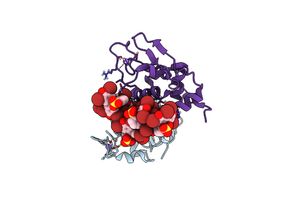
Crystal Structure Of The C-Src Sh3 Domain H122R-Q128K Mutant In Complex With Ni(Ii) At Ph 7.5 Co-Crystallized With Methyl Beta-Cyclodextrin
Organism: Gallus gallus
Method: X-RAY DIFFRACTION Resolution:1.30 Å Release Date: 2020-05-27 Classification: PROTEIN BINDING Ligands: NI |
 |
Organism: Gallus gallus
Method: X-RAY DIFFRACTION Resolution:0.90 Å Release Date: 2020-04-22 Classification: PROTEIN BINDING Ligands: GOL |