
Search Count: 10
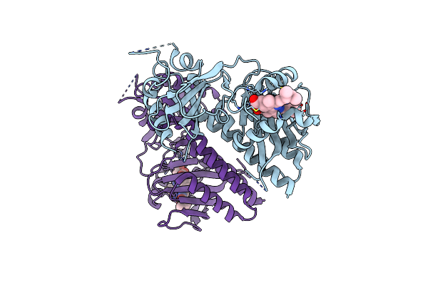 |
Organism: Glycine max
Method: X-RAY DIFFRACTION Resolution:2.20 Å Release Date: 2025-06-04 Classification: PLANT PROTEIN Ligands: CYC |
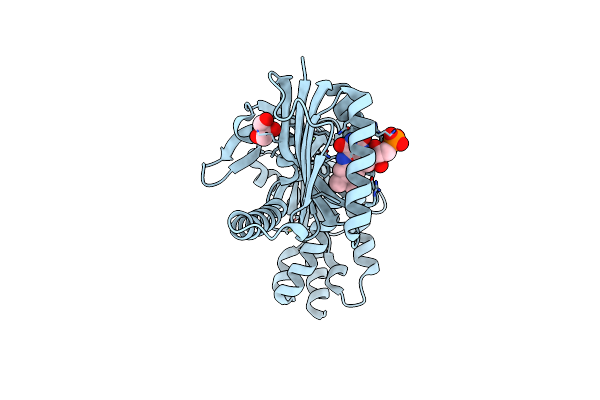 |
Organism: Oscillatoria acuminata
Method: X-RAY DIFFRACTION Resolution:1.95 Å Release Date: 2025-02-26 Classification: LYASE Ligands: FMN, GOL, CA |
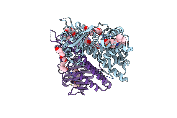 |
Organism: Glycine max
Method: X-RAY DIFFRACTION Resolution:1.58 Å Release Date: 2025-01-08 Classification: PLANT PROTEIN Ligands: CYC, 1PE, PEG, PGE |
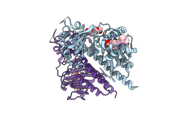 |
Organism: Glycine max
Method: X-RAY DIFFRACTION Resolution:1.86 Å Release Date: 2025-01-08 Classification: PLANT PROTEIN Ligands: O6E, PG4 |
 |
Organism: Glycine max
Method: X-RAY DIFFRACTION Resolution:2.20 Å Release Date: 2025-01-08 Classification: PLANT PROTEIN Ligands: CYC |
 |
Organism: Glycine max
Method: X-RAY DIFFRACTION Resolution:2.20 Å Release Date: 2025-01-08 Classification: PLANT PROTEIN Ligands: CYC |
 |
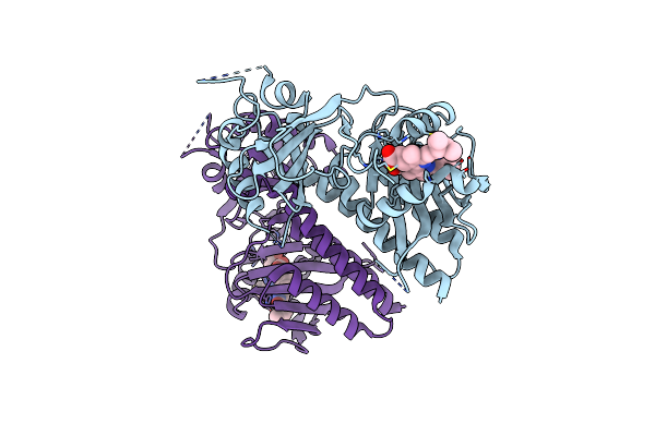
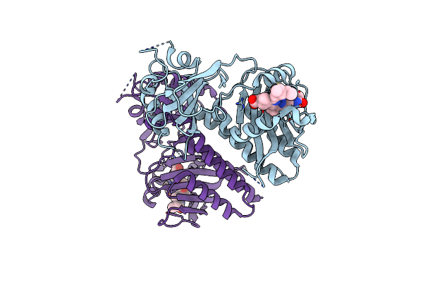
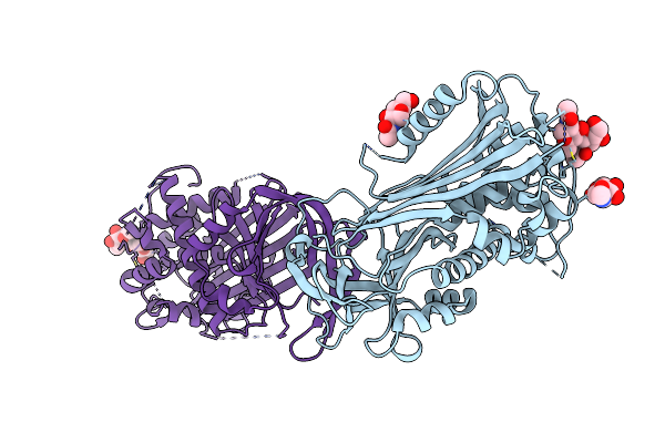
Crystal Structure Of The C. Elegans Pofut2 (Cepofut2) Triple Mutant (R298K-R299K-A418C) In Complex With The Rattus Norvegicus Tsr4 Single Mutant (E10C) From F-Spondin
Organism: Caenorhabditis elegans
Method: X-RAY DIFFRACTION Resolution:2.13 Å Release Date: 2022-10-26 Classification: TRANSFERASE Ligands: NAG, EDO, SO4, GOL |
 |
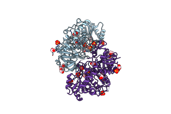
The Structure Of Agmatinase From E. Coli At 1.8 A Displaying Urea And Agmatine
Organism: Escherichia coli
Method: X-RAY DIFFRACTION Resolution:1.80 Å Release Date: 2021-05-12 Classification: HYDROLASE Ligands: AG2, MN, URE, TRS |
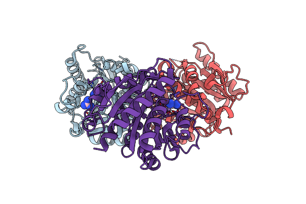 |
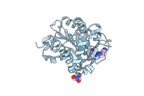
The Structure Of Agmatinase From E. Coli At 3.2 A Displaying Guanidine In The Active Site
Organism: Escherichia coli
Method: X-RAY DIFFRACTION Resolution:3.20 Å Release Date: 2021-05-12 Classification: HYDROLASE Ligands: MN, GAI |
 |
Organism: Homo sapiens
Method: X-RAY DIFFRACTION Resolution:2.80 Å Release Date: 2012-07-18 Classification: BLOOD CLOTTING Ligands: NAG |