
Search Count: 31
 |
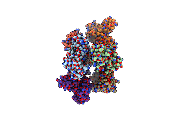
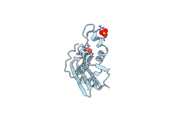
Organism: Lama glama, Severe acute respiratory syndrome coronavirus 2
Method: X-RAY DIFFRACTION Release Date: 2025-05-21 Classification: ANTIVIRAL PROTEIN Ligands: CL |
 |
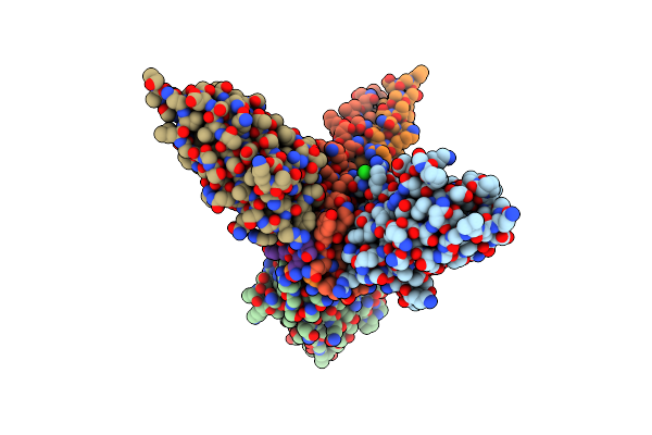
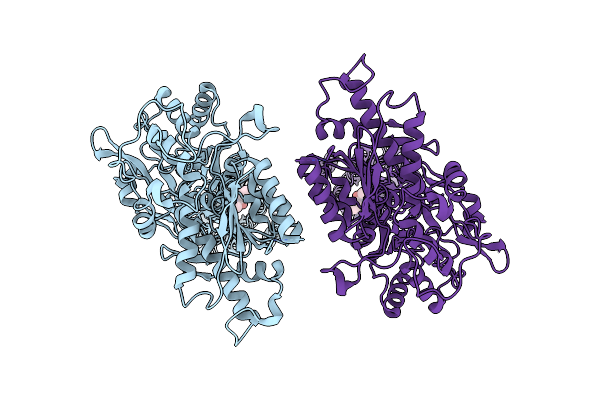
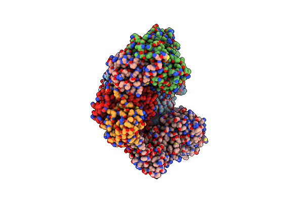
Organism: Homo sapiens
Method: ELECTRON MICROSCOPY Release Date: 2024-11-06 Classification: HYDROLASE/PROTEIN BINDING Ligands: ADP, MG, ATP, ZN |
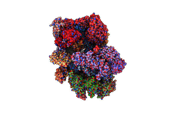 |
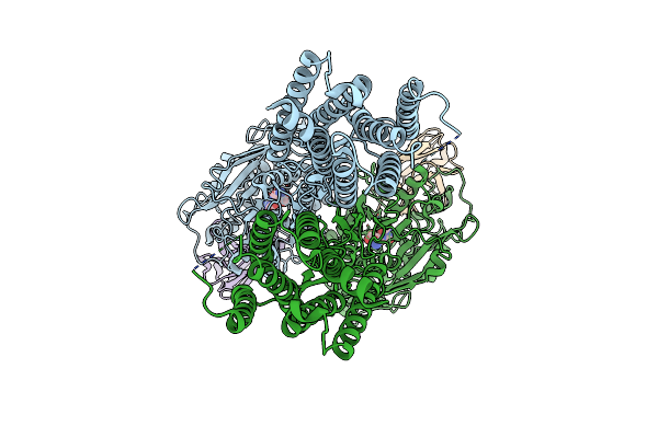
Organism: Homo sapiens
Method: ELECTRON MICROSCOPY Release Date: 2024-11-06 Classification: HYDROLASE/PROTEIN BINDING Ligands: MG, ATP, ADP, ZN |
 |
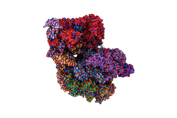
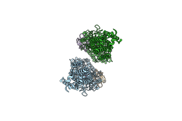
Rpn1/Nub1Ubl-Focused Alignment Of The Non-Substrate-Engaged Human 26S Proteasome
Organism: Homo sapiens
Method: ELECTRON MICROSCOPY Release Date: 2024-11-06 Classification: HYDROLASE/PROTEIN BINDING |
 |
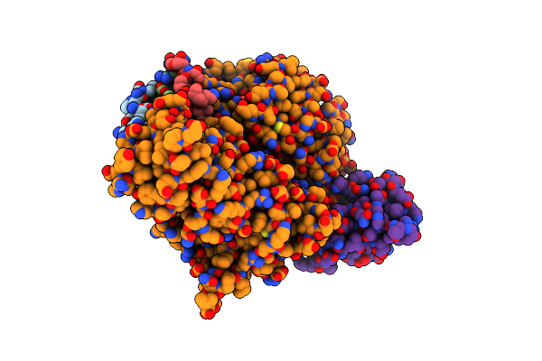
Organism: Homo sapiens
Method: ELECTRON MICROSCOPY Release Date: 2023-12-06 Classification: TRANSLATION |
 |
Eukaryotic Translation Initiation Factor 2B With A Mutation (L516A) In The Delta Subunit
Organism: Homo sapiens
Method: ELECTRON MICROSCOPY Release Date: 2023-12-06 Classification: TRANSLATION |
 |
Organism: Homo sapiens
Method: ELECTRON MICROSCOPY Release Date: 2023-10-11 Classification: SIGNALING PROTEIN Ligands: YKU |
 |
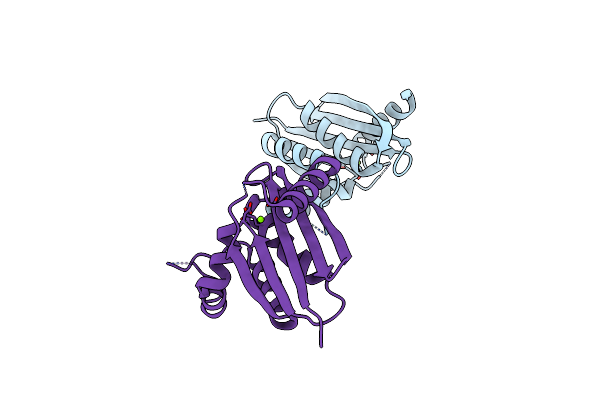
Organism: Homo sapiens, Lama glama
Method: ELECTRON MICROSCOPY Release Date: 2023-10-11 Classification: SIGNALING PROTEIN Ligands: QUS |
 |
Organism: Homo sapiens, Lama glama
Method: ELECTRON MICROSCOPY Release Date: 2023-10-11 Classification: SIGNALING PROTEIN Ligands: QUS |
 |
Organism: Homo sapiens, Lama glama
Method: ELECTRON MICROSCOPY Release Date: 2023-10-11 Classification: SIGNALING PROTEIN Ligands: QUS, YKU |
 |
Organism: Homo sapiens, Lama glama, Synthetic construct
Method: ELECTRON MICROSCOPY Release Date: 2023-04-12 Classification: SIGNALING PROTEIN |
 |
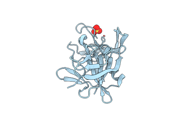
Evolution Avoids A Pathological Stabilizing Interaction In The Immune Protein S100A9
Organism: Homo sapiens
Method: SOLUTION NMR Release Date: 2022-10-26 Classification: SIGNALING PROTEIN Ligands: CA |
 |
Organism: Homo sapiens
Method: X-RAY DIFFRACTION Resolution:3.20 Å Release Date: 2021-10-13 Classification: TRANSFERASE |
 |
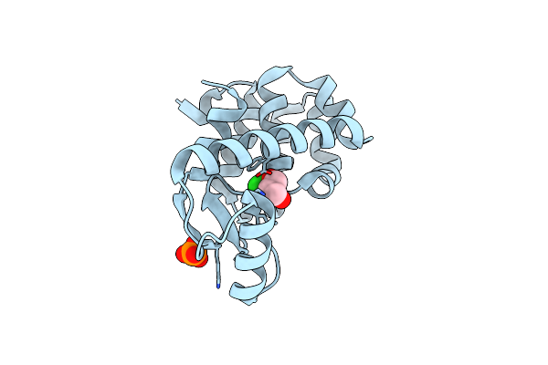
Organism: Streptomyces monomycini
Method: X-RAY DIFFRACTION Resolution:1.25 Å Release Date: 2021-01-20 Classification: UNKNOWN FUNCTION Ligands: SO4 |
 |
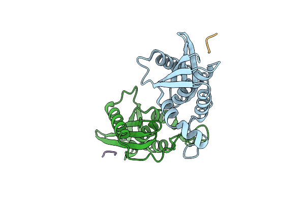
Rubisco / Csos2 N-Peptide Complex Responsible For Alpha-Carboxysome Cargo Loading
Organism: Halothiobacillus neapolitanus (strain atcc 23641 / c2), Halothiobacillus neapolitanus c2
Method: X-RAY DIFFRACTION Resolution:2.40 Å Release Date: 2019-10-30 Classification: PROTEIN BINDING |
 |
Organism: Escherichia coli o139:h28, Escherichia coli
Method: X-RAY DIFFRACTION Resolution:2.00 Å Release Date: 2015-04-29 Classification: Hydrolase/Peptide |
 |
Organism: Unidentified
Method: X-RAY DIFFRACTION Resolution:1.36 Å Release Date: 2014-11-19 Classification: HYDROLASE Ligands: SO4 |
 |
Organism: Chlorobaculum tepidum
Method: X-RAY DIFFRACTION Resolution:1.60 Å Release Date: 2009-05-12 Classification: HYDROLASE Ligands: MG |
 |
Organism: Enterobacteria phage t4
Method: X-RAY DIFFRACTION Resolution:1.90 Å Release Date: 2007-04-10 Classification: HYDROLASE Ligands: CL |
 |
Organism: Enterobacteria phage t4
Method: X-RAY DIFFRACTION Resolution:1.80 Å Release Date: 2007-04-10 Classification: HYDROLASE Ligands: PO4, CL, GOL |