
Search Count: 68
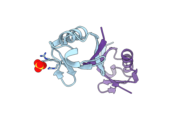 |
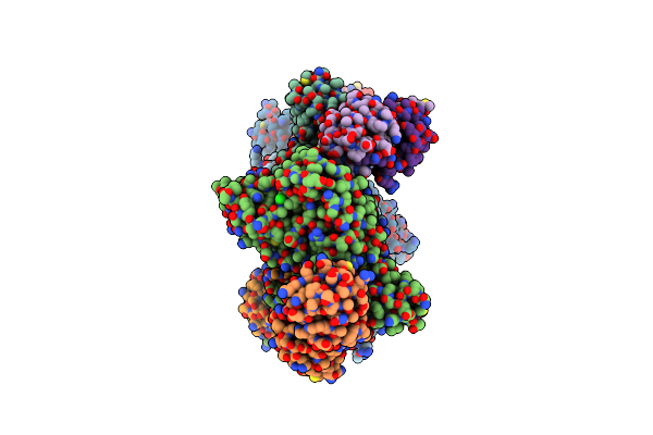
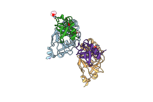
Structure Of A Dimeric Ubiquitin Variant (Ubv3) That Inhibits The Protease (Pro) From Turnip Yellow Mosaic Virus (Tymv)
Organism: Synthetic construct
Method: X-RAY DIFFRACTION Resolution:1.84 Å Release Date: 2025-01-22 Classification: BIOSYNTHETIC PROTEIN Ligands: SO4 |
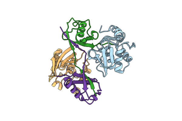 |
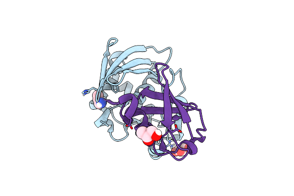
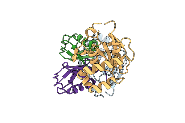
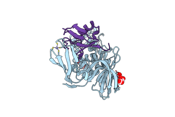
Turnip Yellow Mosaic Virus (Tymv) Protease (Pro) Bound To A Ubiquitin Variant (Ubv3)
Organism: Turnip yellow mosaic virus, Synthetic construct
Method: X-RAY DIFFRACTION Resolution:2.80 Å Release Date: 2025-01-22 Classification: VIRAL PROTEIN |
 |
Organism: Porcine reproductive and respiratory syndrome virus
Method: X-RAY DIFFRACTION Resolution:2.30 Å Release Date: 2023-12-06 Classification: VIRAL PROTEIN Ligands: ZN, ACT |
 |
Organism: Porcine reproductive and respiratory syndrome virus, Homo sapiens
Method: X-RAY DIFFRACTION Resolution:2.85 Å Release Date: 2023-12-06 Classification: VIRAL PROTEIN Ligands: ZN, GOL, 3CN, NO3 |
 |
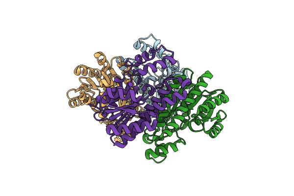
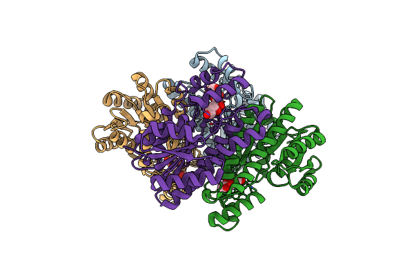
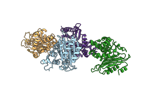
Structure Of The Sars-Cov2 Plpro (C111S) In Complex With A Dimeric Ubv That Inhibits Activity By An Unusual Allosteric Mechanism
Organism: Severe acute respiratory syndrome coronavirus 2, Homo sapiens
Method: X-RAY DIFFRACTION Resolution:3.50 Å Release Date: 2023-01-25 Classification: VIRAL PROTEIN/SIGNALING PROTEIN Ligands: ZN, BR, NA, CL |
 |
Organism: Maize rayado fino virus
Method: X-RAY DIFFRACTION Resolution:1.90 Å Release Date: 2021-07-21 Classification: VIRAL PROTEIN, HYDROLASE |
 |
Organism: Maize rayado fino virus, Homo sapiens
Method: X-RAY DIFFRACTION Resolution:2.09 Å Release Date: 2021-07-21 Classification: VIRAL PROTEIN,HYDROLASE/SUBSTRATE Ligands: GOL, 3CN |
 |
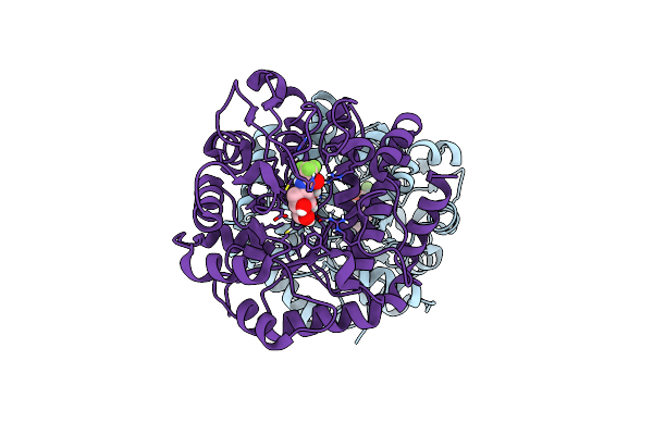
Organism: Sinorhizobium meliloti 1021
Method: X-RAY DIFFRACTION Release Date: 2020-06-24 Classification: OXIDOREDUCTASE |
 |
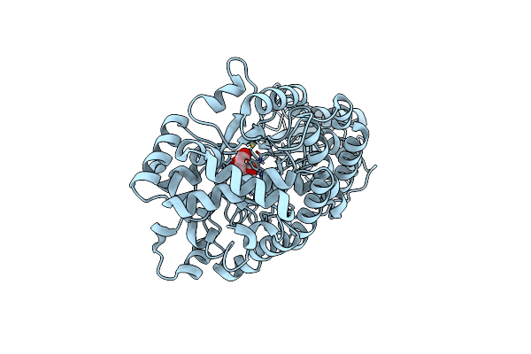
Structure Of Sorbitol Dehydrogenase From Sinorhizobium Meliloti 1021 Bound To Sorbitol
Organism: Sinorhizobium meliloti 1021
Method: X-RAY DIFFRACTION Release Date: 2020-06-24 Classification: OXIDOREDUCTASE Ligands: SOR |
 |
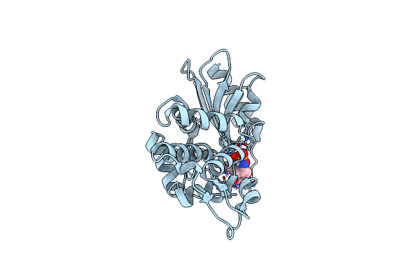
Organism: Burkholderia cenocepacia
Method: X-RAY DIFFRACTION Resolution:1.93 Å Release Date: 2019-04-24 Classification: HYDROLASE Ligands: H9J |
 |
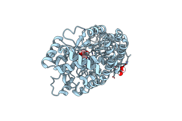
Organism: Vibrio cholerae
Method: X-RAY DIFFRACTION Resolution:1.70 Å Release Date: 2019-01-30 Classification: HYDROLASE |
 |
Organism: Vibrio cholerae
Method: X-RAY DIFFRACTION Resolution:1.90 Å Release Date: 2019-01-30 Classification: HYDROLASE/HYDROLASE INHIBITOR Ligands: NXL |
 |
Organism: Unidentified
Method: X-RAY DIFFRACTION Resolution:2.10 Å Release Date: 2018-01-17 Classification: HYDROLASE Ligands: GOL |
 |
Organism: Unidentified
Method: X-RAY DIFFRACTION Resolution:1.89 Å Release Date: 2018-01-17 Classification: HYDROLASE Ligands: G2F |
 |
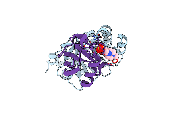
Otu Protease Of Crimean Congo Hemorrhagic Fever Virus Bound To Ubiquitin Variant Cc.4
Organism: Crimean-congo hemorrhagic fever virus (strain nigeria/ibar10200/1970), Synthetic construct
Method: X-RAY DIFFRACTION Resolution:2.10 Å Release Date: 2017-05-10 Classification: hydrolase, transferase Ligands: EDO, NA |
 |
Otu Protease Of Crimean Congo Hemorrhagic Fever Virus Bound To Ubiquitin Variant Cc.2
Organism: Crimean-congo hemorrhagic fever virus (strain nigeria/ibar10200/1970), Synthetic construct
Method: X-RAY DIFFRACTION Resolution:1.50 Å Release Date: 2017-05-10 Classification: hydrolase, transferase |
 |
Otu Protease Of Crimean Congo Hemorrhagic Fever Virus Bound To Ubiquitin Variant Cc.1
Organism: Crimean-congo hemorrhagic fever virus (strain nigeria/ibar10200/1970), Synthetic construct
Method: X-RAY DIFFRACTION Resolution:2.20 Å Release Date: 2017-05-10 Classification: hydrolase, transferase |
 |
Crystal Structure Of The Middle East Respiratory Syndrome Coronavirus Papain-Like Protease Bound To Ubiquitin Variant Me.4
Organism: Human betacoronavirus 2c emc/2012, Homo sapiens
Method: X-RAY DIFFRACTION Resolution:2.55 Å Release Date: 2017-05-10 Classification: HYDROLASE Ligands: ZN, CL, NA, PGO |
 |
Crystal Structure Of The Middle East Respiratory Syndrome Coronavirus Papain-Like Protease Bound To Ubiquitin Variant Me.2
Organism: Human betacoronavirus 2c emc/2012, Homo sapiens
Method: X-RAY DIFFRACTION Resolution:2.70 Å Release Date: 2017-05-10 Classification: HYDROLASE Ligands: ZN, FLC, CL |
 |
Crystal Structure Of Burkholderia Cenocepacia Family 3 Glycoside Hydrolase (Nagz) Bound To N-Ethylbutyryl-Pugnac
Organism: Burkholderia cenocepacia
Method: X-RAY DIFFRACTION Resolution:2.20 Å Release Date: 2017-04-19 Classification: HYDROLASE/HYDROLASE INHIBITOR Ligands: 8M7 |