
Search Count: 12
 |
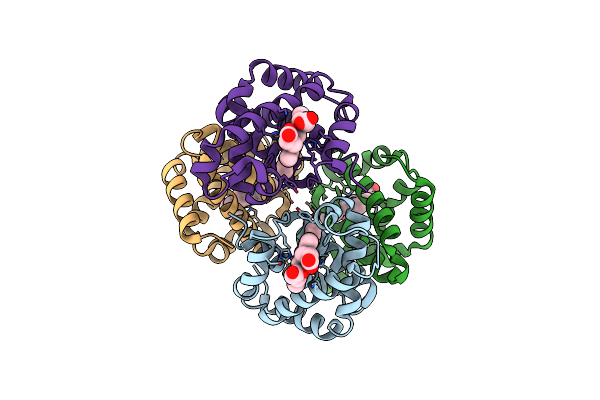
Peanut Lectin Complexed With Divalent N-Beta-D-Galactopyranosyl-L-Tartaramidoyl Derivative (Dingt)
Organism: Arachis hypogaea
Method: X-RAY DIFFRACTION Resolution:1.78 Å Release Date: 2020-10-28 Classification: SUGAR BINDING PROTEIN Ligands: QSG, MN, CA |
 |
Peanut Lectin Complexed With Divalent N-Beta-D-Galactopyranosyl-L-Succinamoyl Derivative (Dings)
Organism: Arachis hypogaea
Method: X-RAY DIFFRACTION Resolution:1.85 Å Release Date: 2020-10-28 Classification: SUGAR BINDING PROTEIN Ligands: QTY, MN, CA |
 |
Peanut Lectin Complexed With N-Beta-D-Galactopyranosyl-L-Succinamoyl Derivative (Ngs)
Organism: Arachis hypogaea
Method: X-RAY DIFFRACTION Resolution:1.75 Å Release Date: 2020-10-28 Classification: SUGAR BINDING PROTEIN Ligands: S3W, MN, CA |
 |
Peanut Lectin Complexed With S-Beta-D-Thiogalactopyranosyl 6-Deoxy-6-S-Propynyl-Beta-D-Glucopyranoside (Stg)
Organism: Arachis hypogaea
Method: X-RAY DIFFRACTION Resolution:1.95 Å Release Date: 2020-10-28 Classification: SUGAR BINDING PROTEIN Ligands: QWJ, MN, CA |
 |
Peanut Lectin Complexed With S-Beta-D-Thiogalactopyranosyl Beta-D-Glucopyranoside Derivative (Stgd)
Organism: Arachis hypogaea
Method: X-RAY DIFFRACTION Resolution:1.90 Å Release Date: 2020-10-28 Classification: SUGAR BINDING PROTEIN Ligands: QWG, MN, CA |
 |
Peanut Lectin Complexed With Divalent S-Beta-D-Thiogalactopyranosyl Beta-D-Glucopyranoside Derivative (Distgd)
Organism: Arachis hypogaea
Method: X-RAY DIFFRACTION Resolution:1.83 Å Release Date: 2020-10-28 Classification: SUGAR BINDING PROTEIN Ligands: WA3, MN, CA |
 |
Genetic And Structural Validation Of Aspergillus Fumigatus N- Acetylphosphoglucosamine Mutase As An Antifungal Target
Organism: Aspergillus fumigatus
Method: X-RAY DIFFRACTION Resolution:2.35 Å Release Date: 2013-05-01 Classification: ISOMERASE Ligands: MG |
 |
The Crystal Structure Of Hemoglobin I From Trematomus Newnesi In Deoxygenated State Obtained Through An Oxidation/Reduction Cycle In Which Potassium Hexacyanoferrate And Sodium Dithionite Were Alternatively Added
Organism: Trematomus newnesi
Method: X-RAY DIFFRACTION Resolution:2.20 Å Release Date: 2010-07-28 Classification: OXYGEN TRANSPORT Ligands: HEM |
 |
The Crystal Structure Of Hemoglobin I From Trematomus Newnesi In Deoxygenated State
Organism: Trematomus newnesi
Method: X-RAY DIFFRACTION Resolution:2.01 Å Release Date: 2010-07-07 Classification: OXYGEN TRANSPORT Ligands: HEM |
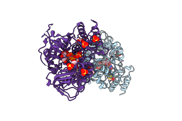 |
Crystal Structure Of Udp-Glucose Phosphorylase From Trypanosoma Brucei, (Tb10.389.0330)
Organism: Trypanosoma brucei
Method: X-RAY DIFFRACTION Resolution:1.92 Å Release Date: 2009-08-18 Classification: TRANSFERASE Ligands: UPG, GOL, SO4, DTU, PG4 |
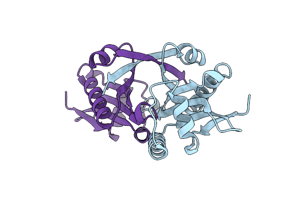 |
Crystal Structure Of Trypanosoma Brucei N-Acetyltransferase (Tb11.01.2886) At 1.86A
Organism: Trypanosoma brucei
Method: X-RAY DIFFRACTION Resolution:1.86 Å Release Date: 2009-08-11 Classification: TRANSFERASE |
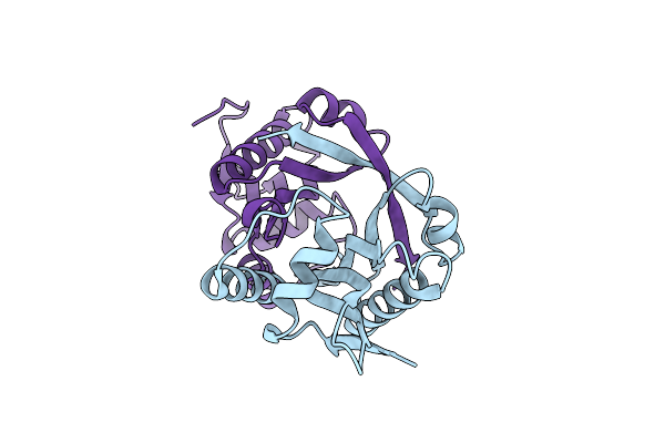 |
Organism: Trypanosoma brucei
Method: X-RAY DIFFRACTION Resolution:2.35 Å Release Date: 2008-11-25 Classification: TRANSFERASE |