
Search Count: 66
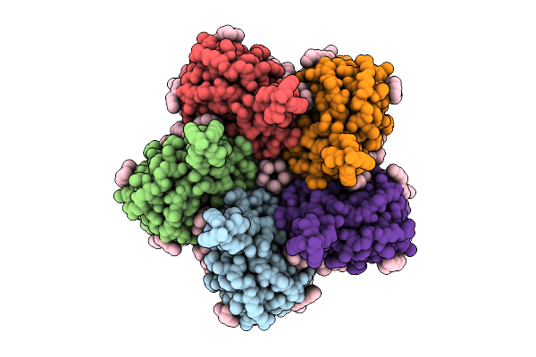 |
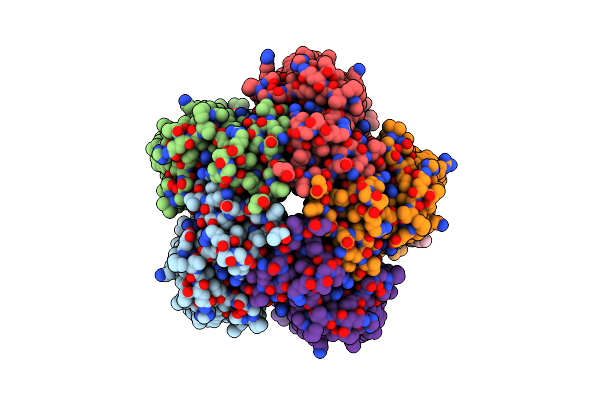
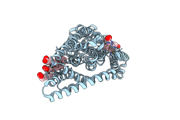
Cryo-Em Structure Of The Flotillin-Associated Rhodopsin Psfar In Detergent Micelle
Organism: Candidatus pseudothioglobus
Method: ELECTRON MICROSCOPY Release Date: 2025-07-23 Classification: MEMBRANE PROTEIN Ligands: LFA |
 |
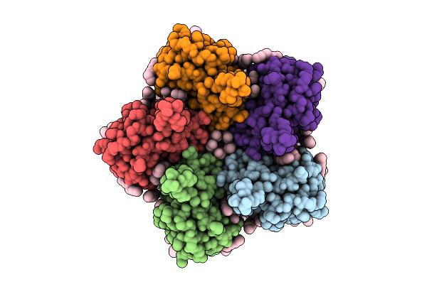
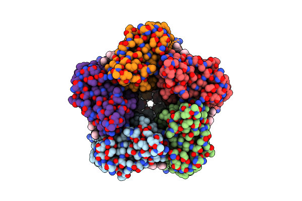
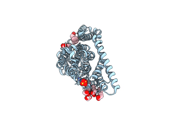
Cryo-Em Structure Of The Light-Driven Proton Pump Pspr In Detergent Micelle
Organism: Candidatus pseudothioglobus sp.
Method: ELECTRON MICROSCOPY Resolution:2.48 Å Release Date: 2025-07-23 Classification: MEMBRANE PROTEIN Ligands: LFA, RET, LMT |
 |
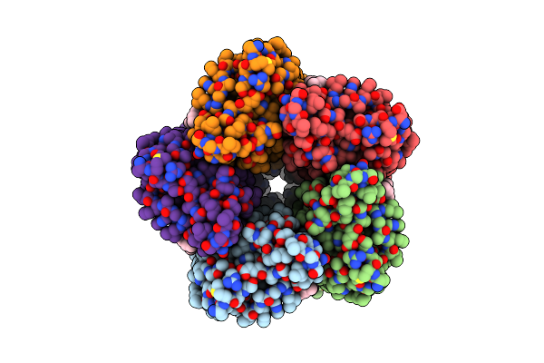
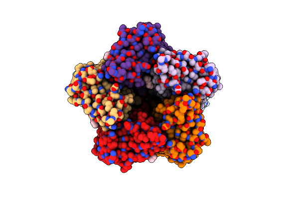
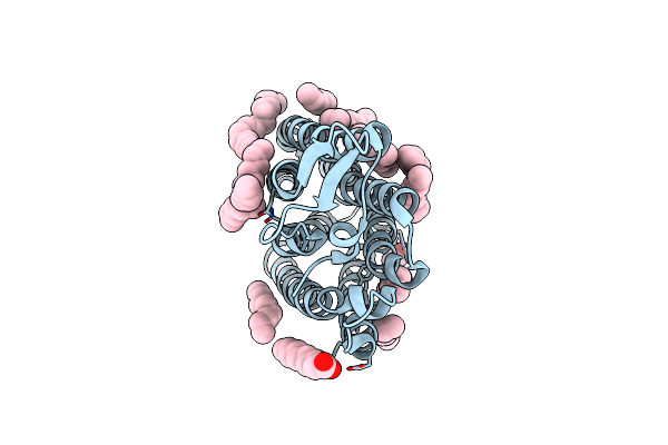
Cryo-Em Structure Of The Double Mutant H84V/E120G Of The Flotillin-Associated Rhodopsin Psfar In Detergent Micelle
Organism: Candidatus pseudothioglobus sp.
Method: ELECTRON MICROSCOPY Resolution:2.81 Å Release Date: 2025-07-23 Classification: MEMBRANE PROTEIN Ligands: LFA, RET |
 |
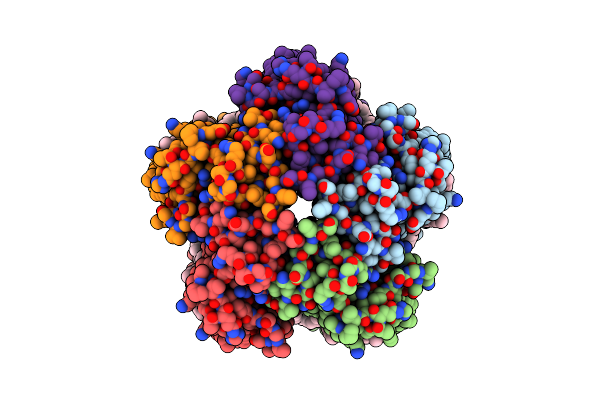
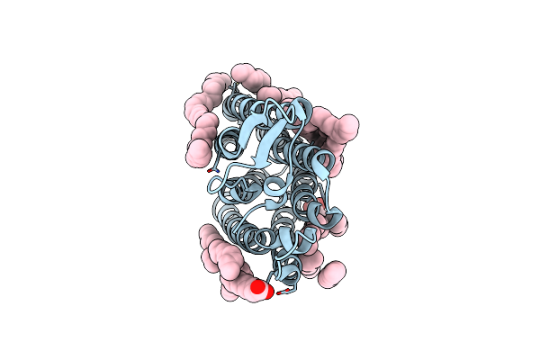
Organism: Cryobacterium levicorallinum
Method: ELECTRON MICROSCOPY Release Date: 2025-05-14 Classification: MEMBRANE PROTEIN Ligands: LFA, RET |
 |
Organism: Cryobacterium levicorallinum
Method: ELECTRON MICROSCOPY Release Date: 2025-05-14 Classification: MEMBRANE PROTEIN Ligands: LFA, RET |
 |
Organism: Cryobacterium levicorallinum
Method: ELECTRON MICROSCOPY Resolution:2.87 Å Release Date: 2025-05-14 Classification: MEMBRANE PROTEIN Ligands: LMT, LFA, RET |
 |
Cryo-Em Structure Of The Microbial Rhodopsin Cryor1 At Ph 10.5 In Detergent In The Ground State
Organism: Cryobacterium levicorallinum
Method: ELECTRON MICROSCOPY Release Date: 2025-05-14 Classification: MEMBRANE PROTEIN Ligands: LMT, RET, LFA |
 |
Cryo-Em Structure Of The Microbial Rhodopsin Cryor1 At Ph 10.5 In Detergent In The M State
Organism: Cryobacterium levicorallinum
Method: ELECTRON MICROSCOPY Release Date: 2025-05-14 Classification: MEMBRANE PROTEIN Ligands: LMT, RET |
 |
Organism: Subtercola endophyticus
Method: ELECTRON MICROSCOPY Resolution:2.44 Å Release Date: 2025-05-14 Classification: MEMBRANE PROTEIN Ligands: LFA, RET |
 |
Organism: Subtercola endophyticus
Method: X-RAY DIFFRACTION Resolution:2.46 Å Release Date: 2024-12-11 Classification: MEMBRANE PROTEIN Ligands: LFA, PO4, RET, OLA |
 |
Crystal Structure Of The Cryorhodopsin Cryor2 At Ph 4.6, Type B Crystals, Non-Illuminated State
Organism: Subtercola sp.
Method: X-RAY DIFFRACTION Resolution:3.00 Å Release Date: 2024-12-11 Classification: MEMBRANE PROTEIN Ligands: LFA, PO4, RET, OLA |
 |
Crystal Structure Of The Cryorhodopsin Cryor2 At Ph 4.6, Type B Crystals, Illuminated State
Organism: Subtercola sp.
Method: X-RAY DIFFRACTION Resolution:3.00 Å Release Date: 2024-12-11 Classification: MEMBRANE PROTEIN Ligands: LFA, PO4, RET, OLA |
 |
Organism: Homo sapiens
Method: X-RAY DIFFRACTION Resolution:2.50 Å Release Date: 2024-11-06 Classification: IMMUNE SYSTEM Ligands: GOL, PGE |
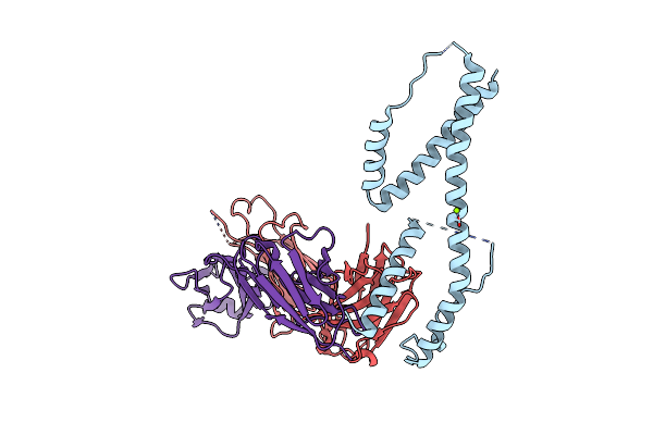 |
Crystal Structure Of The Fab Fragment Of The Anti-Il-6 Antibody I9H In Complex With A Domain-Swapped Il-6 Dimer
Organism: Homo sapiens
Method: X-RAY DIFFRACTION Resolution:2.51 Å Release Date: 2024-11-06 Classification: IMMUNE SYSTEM Ligands: MG |
 |
Crystal Structure Of The Fab Fragment Of The Anti-Il-6 Antibody 68F2 In Complex With A Domain-Swapped Il-6 Dimer
Organism: Homo sapiens, Lama glama
Method: X-RAY DIFFRACTION Resolution:2.93 Å Release Date: 2024-11-06 Classification: IMMUNE SYSTEM Ligands: SO4 |
 |
Crystal Structure Of Apo-Glttk Obtained With In Meso Crystallization (H32 Space Group)
Organism: Thermococcus kodakarensis kod1
Method: X-RAY DIFFRACTION Resolution:2.70 Å Release Date: 2024-09-11 Classification: MEMBRANE PROTEIN Ligands: OLA |
 |
Crystal Structure Of Apo-Glttk Obtained With In Meso Crystallization (P6322 Space Group)
Organism: Thermococcus kodakarensis kod1
Method: X-RAY DIFFRACTION Resolution:3.20 Å Release Date: 2024-09-11 Classification: MEMBRANE PROTEIN Ligands: OLA, OLC, SO4 |
 |
Crystal Structure Of The Light-Driven Sodium Pump Ernar In The Monomeric Form At Ph 4.6
Organism: Erythrobacter
Method: X-RAY DIFFRACTION Resolution:1.70 Å Release Date: 2024-04-24 Classification: MEMBRANE PROTEIN Ligands: LFA, OLA |
 |
Crystal Structure Of The Light-Driven Sodium Pump Ernar In The Monomeric Form At Ph 8.8
Organism: Erythrobacter
Method: X-RAY DIFFRACTION Resolution:1.71 Å Release Date: 2024-04-24 Classification: MEMBRANE PROTEIN Ligands: LFA, OLA |
 |
Cryo-Em Structure Of The Light-Driven Sodium Pump Ernar In The Pentameric Form At Ph 8.0
Organism: Erythrobacter
Method: ELECTRON MICROSCOPY Resolution:2.63 Å Release Date: 2024-04-24 Classification: MEMBRANE PROTEIN Ligands: LFA, LMT |