
Search Count: 4
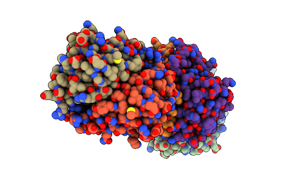 |
Crystal Structure Of The H99N Mutant Of Ribose-5-Phosphate Isomerase B From E. Coli Soaked With Ribose 5-Phosphate
Organism: Escherichia coli
Method: X-RAY DIFFRACTION Resolution:2.10 Å Release Date: 2008-07-08 Classification: ISOMERASE |
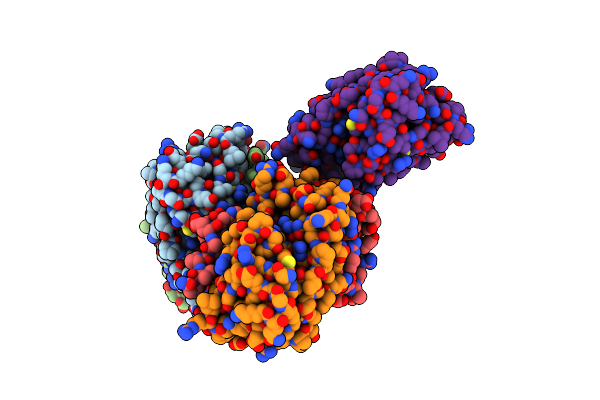 |
Crystal Structure Of Mycobacterium Tuberculosis Ribose-5-Phosphate Isomerase B In Complex With Alpha D-Allose 6-Phosphate
Organism: Mycobacterium tuberculosis
Method: X-RAY DIFFRACTION Resolution:1.85 Å Release Date: 2008-07-01 Classification: ISOMERASE Ligands: A6P |
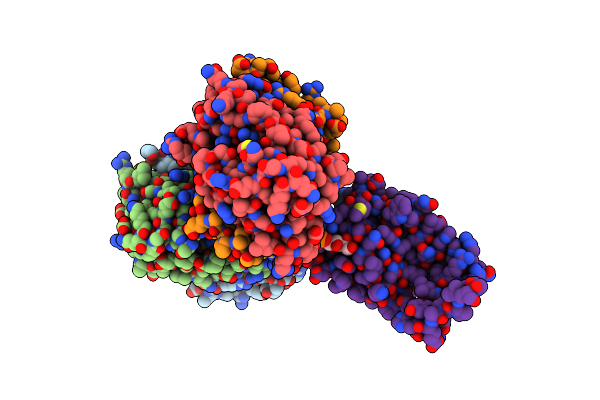 |
Crystal Structure Of Mycobacterium Tuberculosis Ribose-5-Phosphate Isomerase B In Complex With Its Substrates Ribose 5-Phosphate And Ribulose 5-Phosphate
Organism: Mycobacterium tuberculosis
Method: X-RAY DIFFRACTION Resolution:1.65 Å Release Date: 2008-07-01 Classification: ISOMERASE Ligands: R52, 5RP |
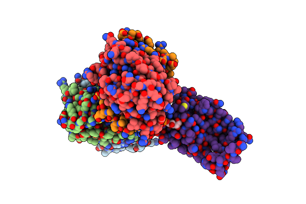 |
Crystal Structure Of Mycobacterium Tuberculosis Ribose-5-Phosphate Isomerase B In Complex With The Inhibitor 5-Deoxy-5-Phospho-D- Ribonate
Organism: Mycobacterium tuberculosis
Method: X-RAY DIFFRACTION Resolution:2.00 Å Release Date: 2008-07-01 Classification: ISOMERASE Ligands: R10, SO4 |