
Search Count: 21
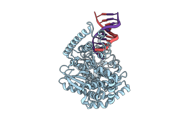 |
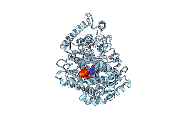
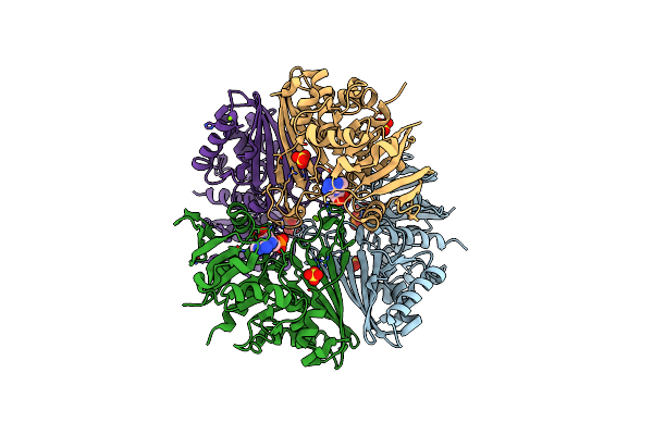
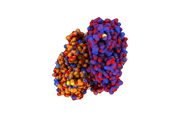
Organism: Plasmodium falciparum, Dna molecule
Method: ELECTRON MICROSCOPY Release Date: 2025-10-29 Classification: REPLICATION |
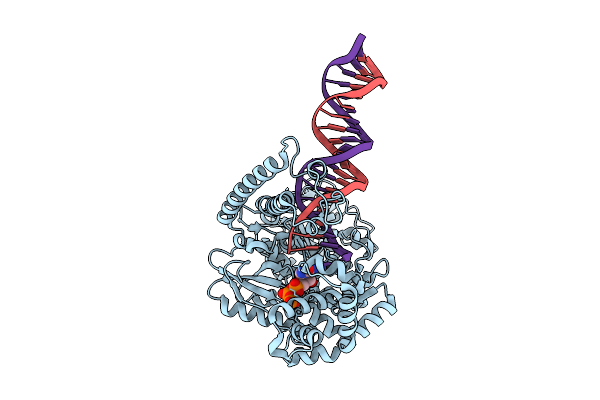 |
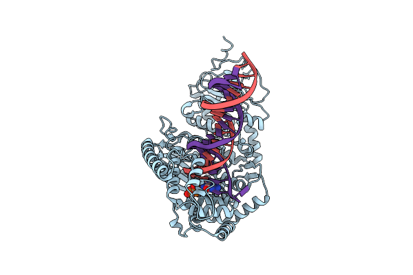
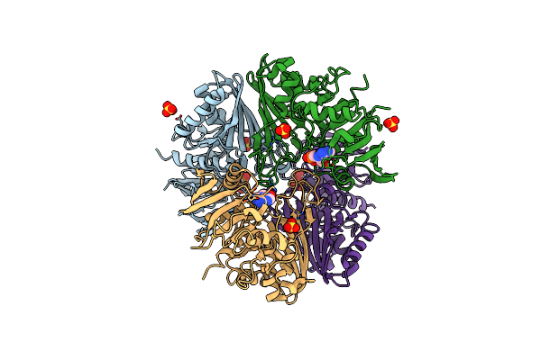
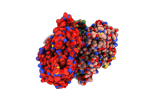
Organism: Plasmodium falciparum, Dna molecule
Method: ELECTRON MICROSCOPY Release Date: 2025-10-29 Classification: REPLICATION Ligands: DGT |
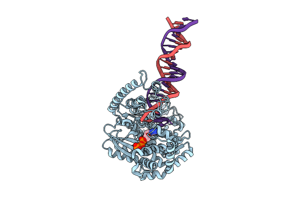 |
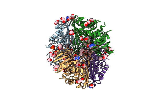
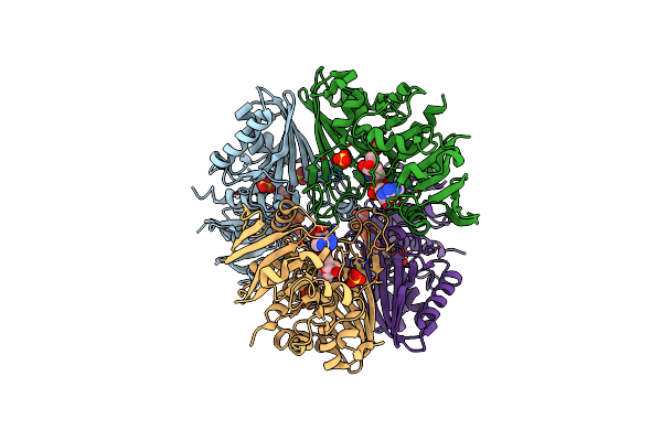
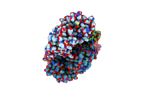
Organism: Plasmodium falciparum, Dna molecule
Method: ELECTRON MICROSCOPY Release Date: 2025-10-29 Classification: REPLICATION Ligands: DGT |
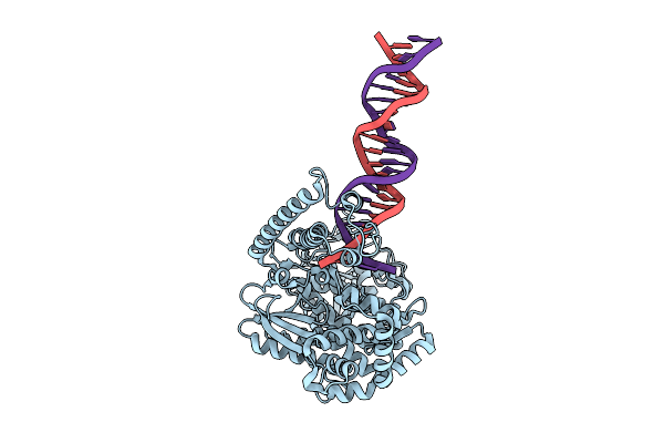 |
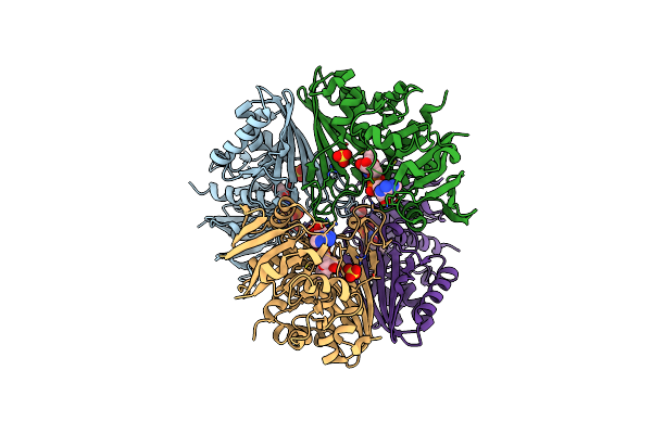
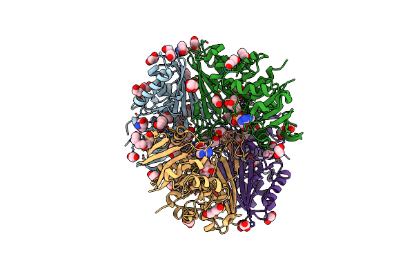
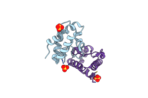
Organism: Plasmodium falciparum, Dna molecule
Method: ELECTRON MICROSCOPY Release Date: 2025-10-29 Classification: REPLICATION |
 |
Organism: Plasmodium falciparum
Method: ELECTRON MICROSCOPY Release Date: 2025-10-29 Classification: REPLICATION Ligands: DGT |
 |
Organism: Plasmodium falciparum, Dna molecule
Method: ELECTRON MICROSCOPY Release Date: 2025-10-29 Classification: REPLICATION Ligands: DGT, CA |
 |
Structure Of The Complex Of Erythrose-4-Phosphate Dehydrogenase From Acinetobacter Baumannii With Nicotinamide Adenine Dinucleotide In The Presence Of Poly(Ethylene Glycol) At 2.20 A Resolution
Organism: Acinetobacter baumannii
Method: X-RAY DIFFRACTION Resolution:2.20 Å Release Date: 2024-07-03 Classification: OXIDOREDUCTASE Ligands: NAD, PEG, PG4, EDO, PO4, PGE, TRS, 1PE, GOL |
 |
Structure Of The Complex Of Erythrose-4-Phosphate Dehydrogenase From Acinetobacter Baumannii With Nicotinamide Adenine Dinucleotide At 2.74 A Resolution.
Organism: Acinetobacter baumannii
Method: X-RAY DIFFRACTION Resolution:2.74 Å Release Date: 2024-07-03 Classification: OXIDOREDUCTASE Ligands: NAD, SO4 |
 |
Crystal Structure Of The Complex Of Erythrose-4-Phosphate Dehydrogenase From Acinetobacter Baumannii With Adenosine Phosphate At 2.40 A Resolution.
Organism: Acinetobacter baumannii
Method: X-RAY DIFFRACTION Resolution:2.40 Å Release Date: 2024-07-03 Classification: OXIDOREDUCTASE Ligands: AMP, SO4, MG |
 |
Crystal Structure Of The Complex Of Glyceraldehyde-3-Phosphate Dehydrogenase Of Type B From Acinetobacter Baumannii With Adenosine Monophosphate At 3.20 A Resolution.
Organism: Acinetobacter baumannii
Method: X-RAY DIFFRACTION Resolution:3.20 Å Release Date: 2024-06-12 Classification: OXIDOREDUCTASE Ligands: AMP, SO4 |
 |
Structure Of Erythrose-4-Phosphate Dehydrogenase From Acinetobacter Baumannii At 3.00 A Resolution
Organism: Acinetobacter baumannii
Method: X-RAY DIFFRACTION Resolution:3.00 Å Release Date: 2024-06-05 Classification: OXIDOREDUCTASE Ligands: NAD, SO4 |
 |
Crystal Structure Of Poly(Ethylene Glycol) Stabilized Erythrose-4-Phosphate Dehydrogenase From Acinetobacter Baumannii At 2.30 A Resolution
Organism: Acinetobacter baumannii
Method: X-RAY DIFFRACTION Resolution:2.30 Å Release Date: 2024-06-05 Classification: OXIDOREDUCTASE Ligands: NAD, PEG, PG4, EDO, TRS, GOL, XPE, PGE, SO4, MG |
 |
Organism: Homo sapiens, Gallus gallus
Method: ELECTRON MICROSCOPY Release Date: 2020-05-20 Classification: CONTRACTILE PROTEIN Ligands: MG, ADP |
 |
Organism: Gallus gallus
Method: ELECTRON MICROSCOPY Release Date: 2020-05-20 Classification: CONTRACTILE PROTEIN Ligands: MG, ADP |
 |
Organism: Saccharomyces cerevisiae, Gallus gallus
Method: ELECTRON MICROSCOPY Release Date: 2020-05-20 Classification: CONTRACTILE PROTEIN/PROTEIN BINDING Ligands: MG, ADP |
 |
Organism: Amanita phalloides, Gallus gallus
Method: ELECTRON MICROSCOPY Release Date: 2020-05-20 Classification: CONTRACTILE PROTEIN/PROTEIN BINDING Ligands: MG, ADP |
 |
Organism: Artemisia vulgaris
Method: X-RAY DIFFRACTION Resolution:1.95 Å Release Date: 2019-03-13 Classification: ALLERGEN Ligands: SO4 |
 |
Oxygenase Component Of 3-Nitrotoluene Dioxygenase From Diaphorobacter Sp. Strain Ds2
Organism: Diaphorobacter sp. ds2
Method: X-RAY DIFFRACTION Resolution:2.90 Å Release Date: 2017-04-19 Classification: METAL BINDING PROTEIN Ligands: FE, FES |
 |
Ferredoxin Component Of 3-Nitrotoluene Dioxygenase From Diaphorobacter Sp. Strain Ds2
Organism: Diaphorobacter sp. ds2
Method: X-RAY DIFFRACTION Resolution:2.40 Å Release Date: 2016-07-27 Classification: METAL BINDING PROTEIN Ligands: FES, FE |
 |
Organism: Actinidia deliciosa
Method: X-RAY DIFFRACTION Resolution:2.10 Å Release Date: 2015-07-29 Classification: PLANT PROTEIN |