
Search Count: 26
 |
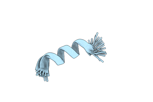
Organism: Klebsiella pneumoniae
Method: X-RAY DIFFRACTION Release Date: 2025-09-03 Classification: VIRAL PROTEIN Ligands: GOL, PEG |
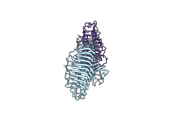 |
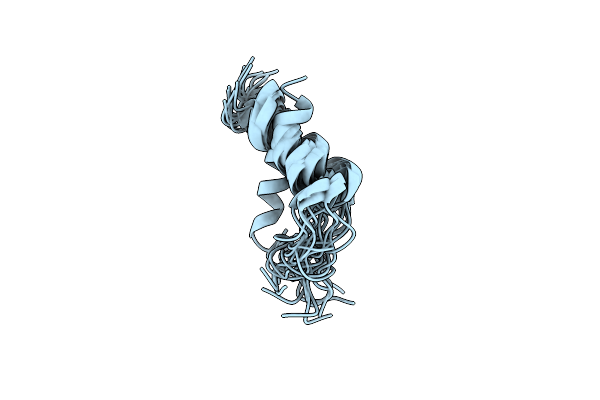
Organism: Klebsiella phage kp34
Method: X-RAY DIFFRACTION Resolution:2.00 Å Release Date: 2023-08-30 Classification: VIRAL PROTEIN |
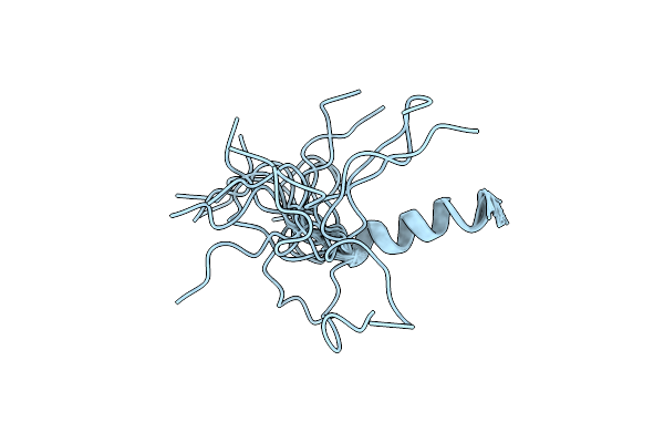 |
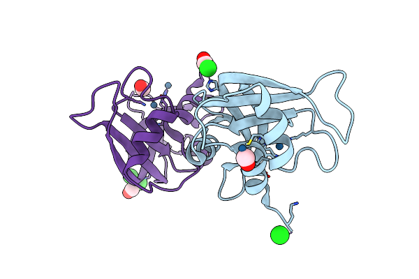
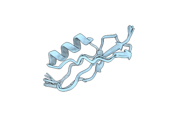
Organism: Homo sapiens
Method: SOLUTION NMR Release Date: 2018-08-01 Classification: SIGNALING PROTEIN |
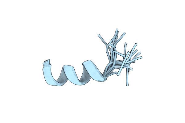 |
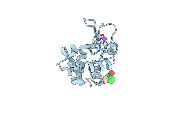
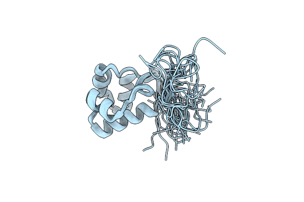
Organism: Homo sapiens
Method: SOLUTION NMR Release Date: 2018-08-01 Classification: SIGNALING PROTEIN |
 |
Organism: Homo sapiens
Method: SOLUTION NMR Release Date: 2018-08-01 Classification: SIGNALING PROTEIN |
 |
Organism: Homo sapiens
Method: SOLUTION NMR Release Date: 2017-12-20 Classification: SIGNALING PROTEIN |
 |
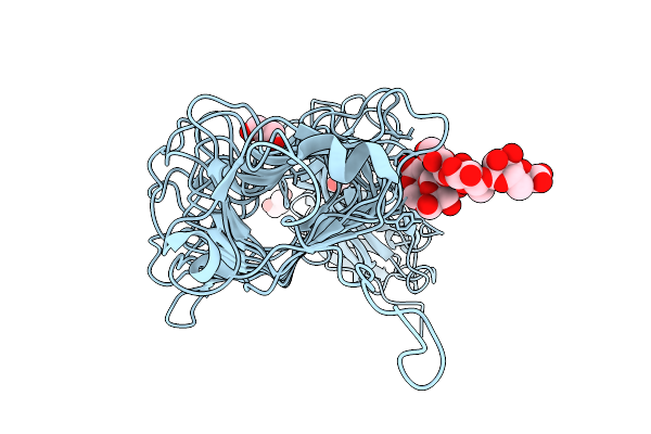
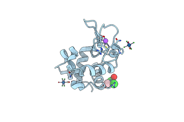
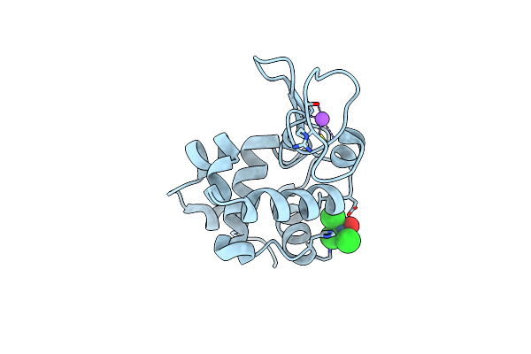
X-Ray Structure Of The Complex Between Bovine Pancreatic Ribonuclease And Pentachlorocarbonyliridate(Iii) (4 Days Of Soaking)
Organism: Bos taurus
Method: X-RAY DIFFRACTION Resolution:1.85 Å Release Date: 2016-07-27 Classification: HYDROLASE Ligands: IR, CL, CMO |
 |
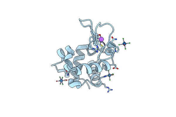
X-Ray Structure Of The Complex Between Bovine Pancreatic Ribonuclease And Penthachlorocarbonyliridate(Iii) (2 Months Of Soaking)
Organism: Bos taurus
Method: X-RAY DIFFRACTION Resolution:2.29 Å Release Date: 2016-07-27 Classification: HYDROLASE Ligands: IR, O, CL, CMO |
 |
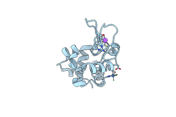
X-Ray Structure Of The Complex Between Hen Egg White Lysozyme And Pentacholrocarbonyliridate(Iii) (1 Day)
Organism: Gallus gallus
Method: X-RAY DIFFRACTION Resolution:1.55 Å Release Date: 2015-12-30 Classification: HYDROLASE Ligands: 2T8, CL, NA |
 |
Organism: Homo sapiens
Method: SOLUTION NMR Release Date: 2015-07-08 Classification: SIGNALING PROTEIN |
 |
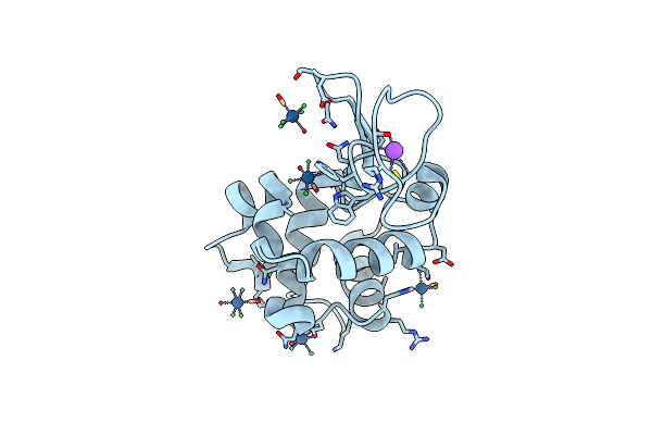
X-Ray Structure Of The Bis-Platinum Lysozyme Adduct Formed In The Reaction Between The Protein And The Two Drugs Cisplatin And Oxaliplatin
Organism: Gallus gallus
Method: X-RAY DIFFRACTION Resolution:1.85 Å Release Date: 2015-05-27 Classification: HYDROLASE Ligands: EDO, NO3, NA, 1PT, CPT |
 |
X-Ray Structure Of The Bis-Platinum Lysozyme Adduct Formed In The Reaction Between The Protein And The Two Drugs Cisplatin And Oxaliplatin (Preparation 2)
Organism: Gallus gallus
Method: X-RAY DIFFRACTION Resolution:1.95 Å Release Date: 2015-05-27 Classification: HYDROLASE Ligands: NO3, EDO, 1PT, CPT |
 |
X-Ray Structure Of The Adduct Between Hen Egg White Lysozyme And Auoxo3, A Cytotoxic Gold(Iii) Compound
Organism: Gallus gallus
Method: X-RAY DIFFRACTION Resolution:2.05 Å Release Date: 2014-11-05 Classification: HYDROLASE Ligands: NO3, EDO, NA, AU |
 |
X-Ray Structure Of The Complex Between The Hen Egg White Lysozyme And Pentachlorocarbonyliridate (Iii) (4 Days)
Organism: Gallus gallus
Method: X-RAY DIFFRACTION Resolution:1.42 Å Release Date: 2014-09-17 Classification: HYDROLASE Ligands: 2T8, CL, NA |
 |
X-Ray Structure Of The Complex Between Hen Egg White Lysozyme And Pentachlorocarbonyliridate(Iii) (5 Days)
Organism: Gallus gallus
Method: X-RAY DIFFRACTION Resolution:1.92 Å Release Date: 2014-09-17 Classification: HYDROLASE Ligands: 2T8, CL, NA |
 |
X-Ray Structure Of The Complex Between Hen Egg White Lysozyme And Pentachlorocarbonyliridate(Iii) (9 Days)
Organism: Gallus gallus
Method: X-RAY DIFFRACTION Resolution:1.99 Å Release Date: 2014-09-17 Classification: HYDROLASE Ligands: 2T8, CL, NA |
 |
X-Ray Structure Of The Complex Between Hen Egg White Lysozyme And Pentachlorocarbonyliridate(Iii) (6 Days)
Organism: Gallus gallus
Method: X-RAY DIFFRACTION Resolution:1.65 Å Release Date: 2014-09-17 Classification: HYDROLASE Ligands: 2T8, CL, NA |
 |
X-Ray Structure Of The Complex Between Hen Egg White Lysozyme And Pentachlorocarbonyliridate(Iii) (30 Days)
Organism: Gallus gallus
Method: X-RAY DIFFRACTION Resolution:1.86 Å Release Date: 2014-09-17 Classification: HYDROLASE Ligands: 2T8, CL, NA |
 |
Organism: Mycobacterium tuberculosis
Method: SOLUTION NMR Release Date: 2013-12-25 Classification: PENICILLIN BINDING PROTEIN |
 |
Organism: Homo sapiens
Method: SOLUTION NMR Release Date: 2012-03-14 Classification: SIGNALING PROTEIN |