
Search Count: 20
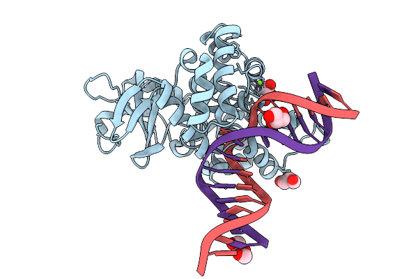 |
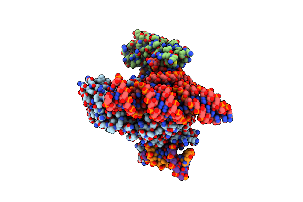
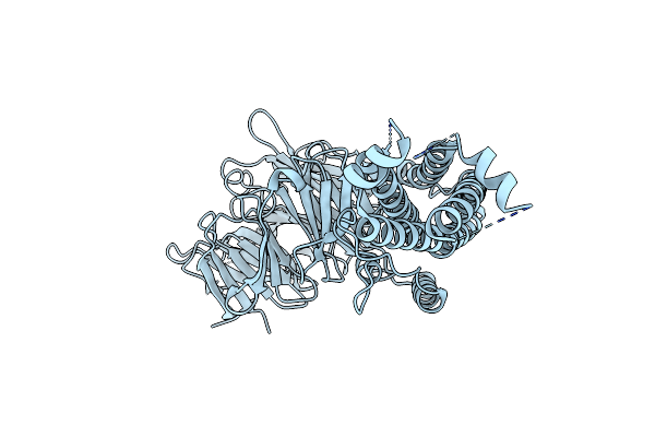
Crystal Structure Of Human 8-Oxoguanine Glycosylase K249H Mutant Bound To The Substrate 8-Oxoguanine Dna At Ph 8.0 Under 277 K
Organism: Homo sapiens
Method: X-RAY DIFFRACTION Resolution:1.45 Å Release Date: 2025-07-23 Classification: DNA/HYDROLASE Ligands: PEG, GOL, MG, NA |
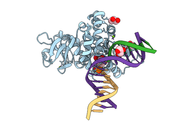 |
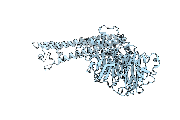
Crystal Structure Of Human 8-Oxoguanine Glycosylase K249H Mutant Bound To The Reaction Intermediate Derived From The Crystal Soaked Into The Solution At Ph 4.0 Under 277 K For 24 Hourss
Organism: Homo sapiens
Method: X-RAY DIFFRACTION Resolution:1.68 Å Release Date: 2025-07-23 Classification: DNA/HYDROLASE Ligands: A1LXK, MG, GOL |
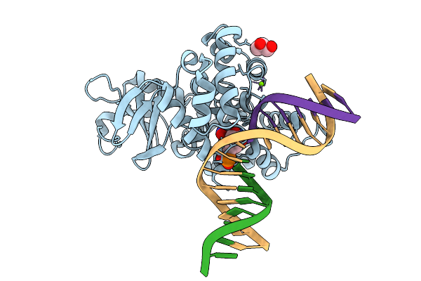 |
Crystal Structure Of Human 8-Oxoguanine Glycosylase K249H Mutant Bound To The Reaction Intermediate Derived From The Crystal Soaked Into The Solution At Ph 4.0 Under 277 K For 2.5 Hours
Organism: Homo sapiens
Method: X-RAY DIFFRACTION Resolution:1.82 Å Release Date: 2025-07-23 Classification: DNA/HYDROLASE Ligands: A1LXK, MG, GOL |
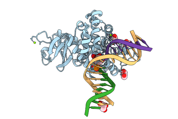 |
Crystal Structure Of Human 8-Oxoguanine Glycosylase K249H Mutant Bound To The Reaction Intermediate Derived From The Crystal Soaked Into The Solution At Ph 4.0 Under 298 K For 3 Weeks
Organism: Homo sapiens
Method: X-RAY DIFFRACTION Resolution:1.70 Å Release Date: 2025-07-23 Classification: DNA/HYDROLASE Ligands: A1LXK, GOL, MG, NA |
 |
Organism: Acidibacillus sulfuroxidans, Acidibacillus sulfooxidans
Method: ELECTRON MICROSCOPY Release Date: 2023-09-27 Classification: RNA BINDING PROTEIN/DNA/RNA Ligands: MG, ZN |
 |
Organism: Sulfoacidibacillus thermotolerans
Method: ELECTRON MICROSCOPY Release Date: 2023-09-27 Classification: RNA BINDING PROTEIN/DNA/RNA Ligands: ZN, MG |
 |
Organism: Sulfoacidibacillus thermotolerans
Method: ELECTRON MICROSCOPY Release Date: 2023-09-27 Classification: RNA BINDING PROTEIN/DNA/RNA Ligands: ZN, MG |
 |
Organism: Homo sapiens
Method: X-RAY DIFFRACTION Resolution:3.20 Å Release Date: 2020-01-15 Classification: TRANSCRIPTION Ligands: P7F |
 |
Crystal Structure Of Vitamin D Hydroxylase Cytochrome P450 105A1 (R84A Mutant) In Complex With 1,25-Dihydroxyvitamin D2
Organism: Streptomyces griseolus
Method: X-RAY DIFFRACTION Resolution:1.90 Å Release Date: 2017-05-10 Classification: OXIDOREDUCTASE Ligands: HEM, 7ZU |
 |
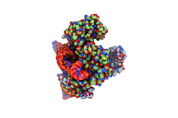
Atomic-Resolution Structures Of The Apc/C Subunits Apc4 And The Apc5 N-Terminal Domain
Organism: Xenopus laevis
Method: X-RAY DIFFRACTION Resolution:3.20 Å Release Date: 2015-09-02 Classification: CELL CYCLE |
 |
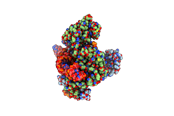
Atomic-Resolution Structures Of The Apc/C Subunits Apc4 And The Apc5 N-Terminal Domain
Organism: Homo sapiens
Method: X-RAY DIFFRACTION Resolution:3.40 Å Release Date: 2015-09-02 Classification: CELL CYCLE |
 |
Atomic-Resolution Structures Of The Apc/C Subunits Apc4 And The Apc5 N-Terminal Domain
Organism: Xenopus laevis
Method: X-RAY DIFFRACTION Resolution:2.18 Å Release Date: 2015-09-02 Classification: CELL CYCLE Ligands: EDO |
 |
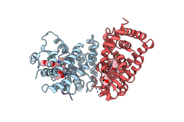
Crystal Structure Of Alcaligenes Faecalis D-3-Hydroxybutyrate Dehydrogenase In Complex With Nad(+) And Acetate
Organism: Alcaligenes faecalis
Method: X-RAY DIFFRACTION Resolution:2.20 Å Release Date: 2012-02-29 Classification: OXIDOREDUCTASE Ligands: ACT, CL, NAD, CA |
 |
Crystal Structure Of D-3-Hydroxybutyrate Dehydrogenase, Prepared In The Presence Of The Substrate D-3-Hydroxybutyrate And Nad(+)
Organism: Alcaligenes faecalis
Method: X-RAY DIFFRACTION Resolution:3.00 Å Release Date: 2012-02-08 Classification: OXIDOREDUCTASE Ligands: CA, CL, NAD, NAI, 3HR, AAE |
 |
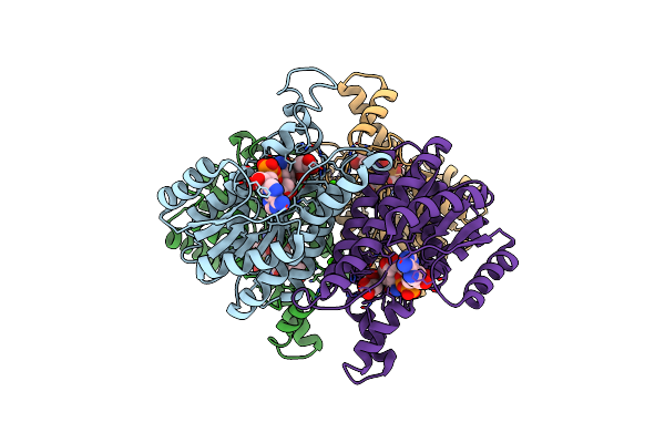
Crystal Structure Of Uricase From Arthrobacter Globiformis In Complex With Uric Acid (Substrate)
Organism: Arthrobacter globiformis
Method: X-RAY DIFFRACTION Resolution:1.90 Å Release Date: 2008-05-06 Classification: OXIDOREDUCTASE Ligands: URC |
 |
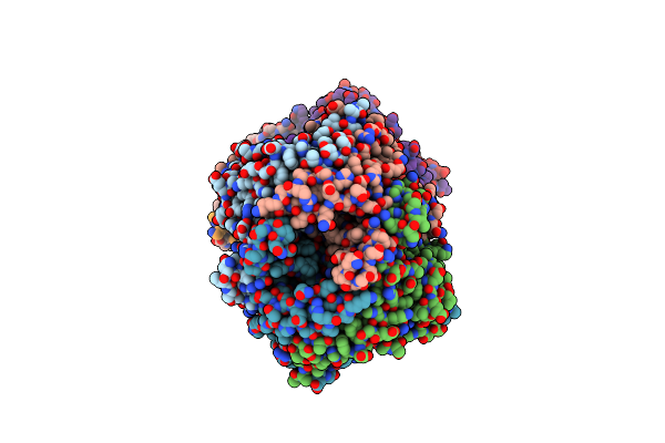
Crystal Structure Of Uricase From Arthrobacter Globiformis In Complex With Allantoate
Organism: Arthrobacter globiformis
Method: X-RAY DIFFRACTION Resolution:1.88 Å Release Date: 2008-05-06 Classification: OXIDOREDUCTASE Ligands: 1AL |
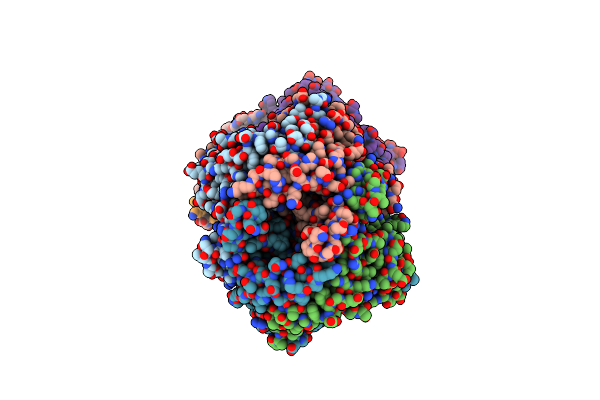 |
Crystal Structure Of Uricase From Arthrobacter Globiformis In Complex With 8-Azaxanthin (Inhibitor)
Organism: Arthrobacter globiformis
Method: X-RAY DIFFRACTION Resolution:2.24 Å Release Date: 2008-05-06 Classification: OXIDOREDUCTASE Ligands: AZA |
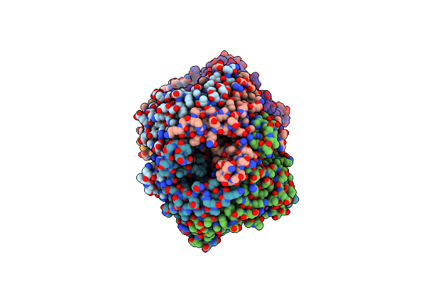 |
Organism: Arthrobacter globiformis
Method: X-RAY DIFFRACTION Resolution:1.99 Å Release Date: 2008-05-06 Classification: OXIDOREDUCTASE Ligands: NOB |
 |
X-Ray Analyses Of 3-Hydroxybutyrate Dehydrogenase From Alcaligenes Faecalis
Organism: Alcaligenes faecalis
Method: X-RAY DIFFRACTION Resolution:2.19 Å Release Date: 2008-04-22 Classification: OXIDOREDUCTASE Ligands: CA, CL |
 |
Organism: Sarcophaga peregrina
Method: SOLUTION NMR Release Date: 2002-03-27 Classification: ANTIBIOTIC |