
Search Count: 610
 |
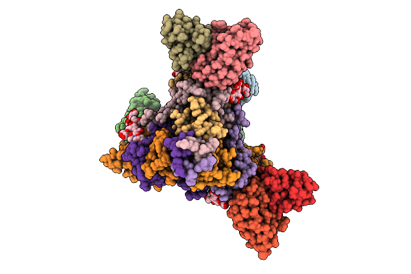
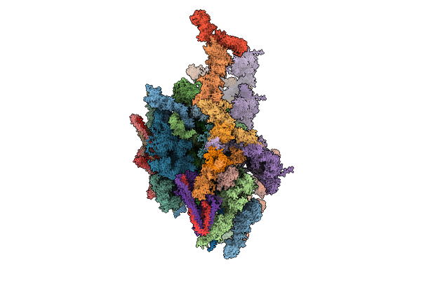
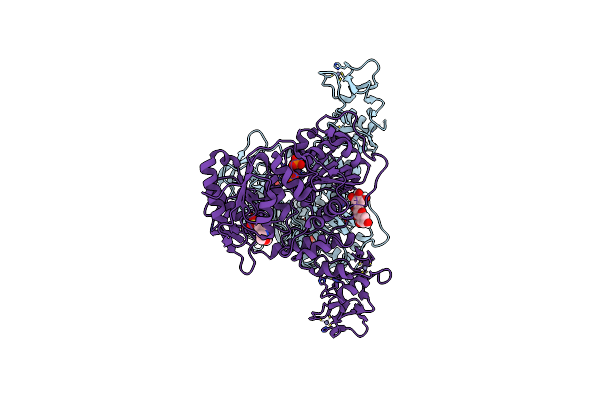
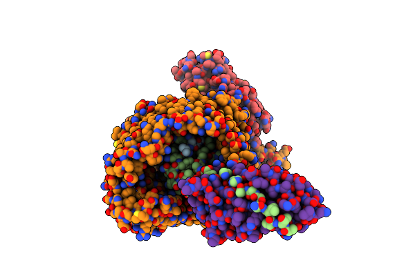
Cryo Em Structure Of Rc-Dlh Complex Model Ii From Gemmatimonas Groenlandica
Organism: Gemmatimonas groenlandica
Method: ELECTRON MICROSCOPY Release Date: 2025-12-03 Classification: PHOTOSYNTHESIS Ligands: BCL, BPH, LMT, MQ8, FE, CD4, CRT, PEX, HEC, V7N |
 |
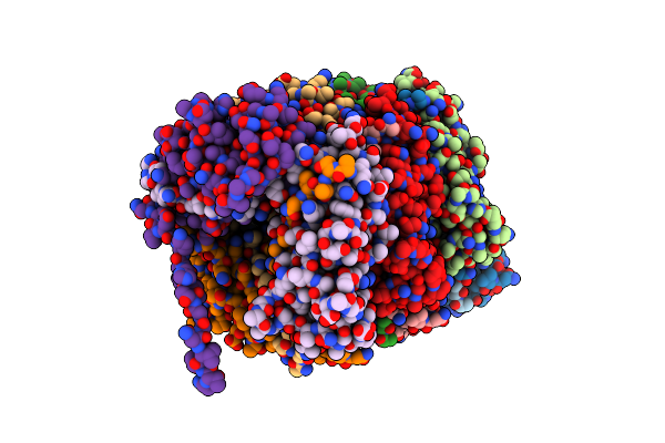
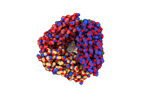
Organism: Homo sapiens, Severe acute respiratory syndrome coronavirus 2
Method: ELECTRON MICROSCOPY Release Date: 2025-11-26 Classification: VIRUS Ligands: NAG |
 |
Organism: Homo sapiens, Severe acute respiratory syndrome coronavirus 2
Method: ELECTRON MICROSCOPY Release Date: 2025-11-26 Classification: VIRUS |
 |
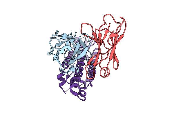
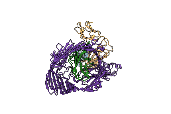
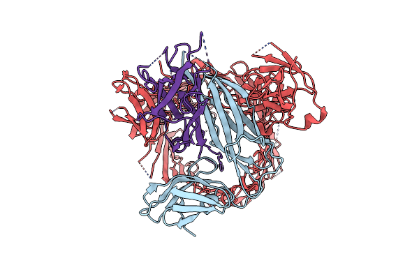
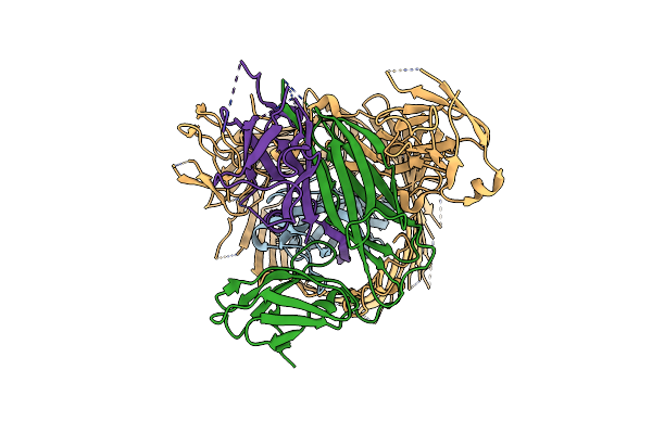
A Broadly-Neutralizing Antibody Against Ebolavirus Glycoprotein That Can Potentiate The Breadth And Neutralization Potency Of Other Anti-Glycoprotein Antibodies
Organism: Oryctolagus cuniculus, Ebolavirus
Method: ELECTRON MICROSCOPY Release Date: 2025-11-05 Classification: VIRAL PROTEIN/IMMUNE SYSTEM Ligands: NAG |
 |
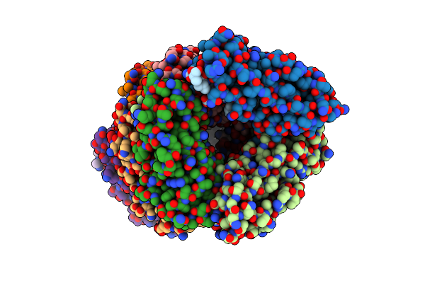
Organism: Arabidopsis thaliana
Method: ELECTRON MICROSCOPY Release Date: 2025-11-05 Classification: NUCLEAR PROTEIN |
 |
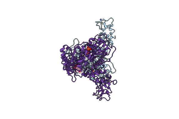
Cryo-Em Structure Of Rc-Dlh Complex Model I From Gem. Groenlandica Strain Tet16
Organism: Gemmatimonas groenlandica
Method: ELECTRON MICROSCOPY Release Date: 2025-10-29 Classification: PHOTOSYNTHESIS Ligands: V7N, BCL, LMT, PEX, MQ8, CD4, BPH, FE, CRT, HEC |
 |
Organism: Homo sapiens
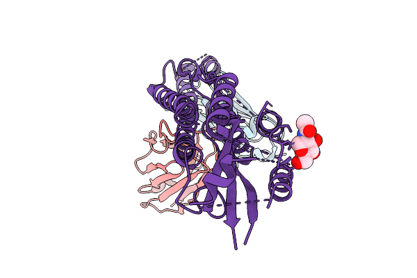
Method: X-RAY DIFFRACTION Release Date: 2025-09-24 Classification: HYDROLASE Ligands: MG, EDO, A1JD4 |
 |
Organism: Homo sapiens
Method: X-RAY DIFFRACTION Release Date: 2025-09-24 Classification: HYDROLASE Ligands: A1JED, TEW, MG |
 |
Organism: Severe acute respiratory syndrome coronavirus 2
Method: X-RAY DIFFRACTION Release Date: 2025-07-30 Classification: HYDROLASE Ligands: ZN, ADP, PO4, MPO |
 |
Organism: Severe acute respiratory syndrome coronavirus 2
Method: X-RAY DIFFRACTION Release Date: 2025-07-23 Classification: HYDROLASE Ligands: ZN, ATP, PO4, MPO |
 |
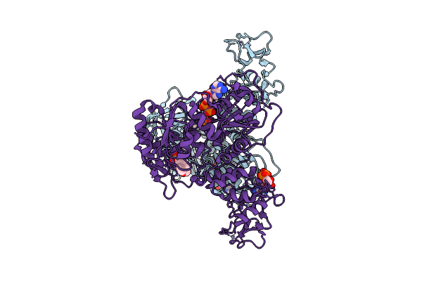
Crystal Structure Of The Sars-Cov-2 Helicase Nsp13 In Complex With Myricetin
Organism: Severe acute respiratory syndrome coronavirus 2
Method: X-RAY DIFFRACTION Release Date: 2025-06-11 Classification: HYDROLASE Ligands: ZN, PO4, MPO, MYC |
 |
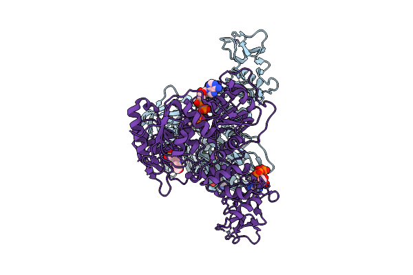
Organism: Severe acute respiratory syndrome coronavirus 2
Method: X-RAY DIFFRACTION Release Date: 2025-06-11 Classification: HYDROLASE Ligands: ZN, PO4, MPO |
 |
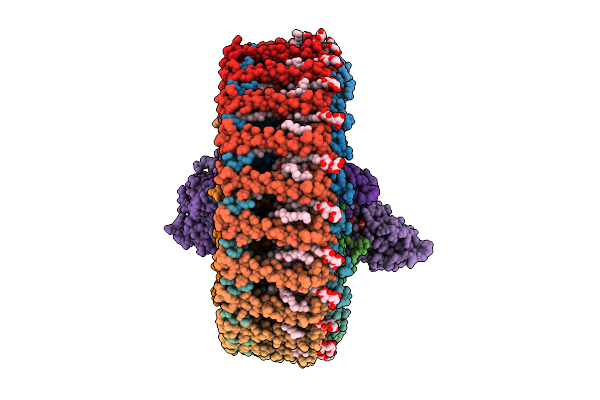
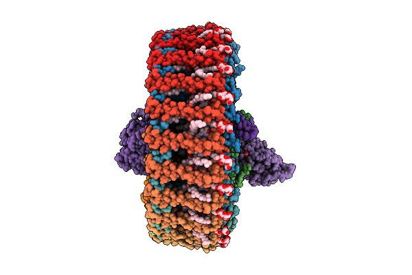
Cryo-Em Structure Of The Type 1 Pilus Rod As Part Of The Fima-Bound Usher Complex (Fimdhgfanc)
Organism: Escherichia coli
Method: ELECTRON MICROSCOPY Release Date: 2025-06-04 Classification: CELL ADHESION |
 |
Cryo-Em Structure Of The Type 1 Pilus Assembly Platform As Part Of The Fima-Bound Chaperone-Usher Pilus Complex (Fimdhgfanc - Body 2)
Organism: Escherichia coli
Method: ELECTRON MICROSCOPY Release Date: 2025-06-04 Classification: MEMBRANE PROTEIN |
 |
Cryo-Em Structure Of The Type 1 Pilus Assembly Platform As Part Of The Fima-Bound Chaperone-Usher Pilus Complex (Local Refinement Including Fimd, Fimc, Fiman And Fiman-1)
Organism: Escherichia coli
Method: ELECTRON MICROSCOPY Release Date: 2025-06-04 Classification: MEMBRANE PROTEIN |
 |
Cryo-Em Structure Of The Type 1 Pilus Assembly Platform As Part Of The Fima-Bound Chaperone-Usher Pilus Complex (Local Refinement Including Fimd, Fimc, Fiman, Fiman-1 And Fiman-2)
Organism: Escherichia coli
Method: ELECTRON MICROSCOPY Release Date: 2025-06-04 Classification: MEMBRANE PROTEIN |
 |
Organism: Escherichia coli
Method: ELECTRON MICROSCOPY Release Date: 2025-06-04 Classification: CELL ADHESION |
 |
Cryo-Em Structure Of The Fimi-Bound Type 1 Pilus Assembly Platform Complex - Local Refinement
Organism: Escherichia coli
Method: ELECTRON MICROSCOPY Release Date: 2025-06-04 Classification: MEMBRANE PROTEIN |
 |
Cryo-Em Structure Of The Type 1 Pilus Rod As Part Of The Fimi-Bound Usher Complex (Fimdhgfanic - Body 1)
Organism: Escherichia coli
Method: ELECTRON MICROSCOPY Release Date: 2025-06-04 Classification: CELL ADHESION |
 |
Cryo-Em Structure Of The Type 1 Pilus Assembly Platform As Part Of The Fimi-Bound Chaperone-Usher Pilus Complex (Fimdhgfanic - Body 2)
Organism: Escherichia coli
Method: ELECTRON MICROSCOPY Release Date: 2025-06-04 Classification: MEMBRANE PROTEIN |