
Search Count: 523
 |
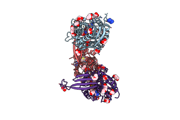
Organism: Streptomyces lividans 1326
Method: X-RAY DIFFRACTION Release Date: 2025-10-08 Classification: OXIDOREDUCTASE Ligands: HEM |
 |
Organism: Rattus norvegicus
Method: ELECTRON MICROSCOPY Release Date: 2025-10-08 Classification: PROTEIN BINDING |
 |
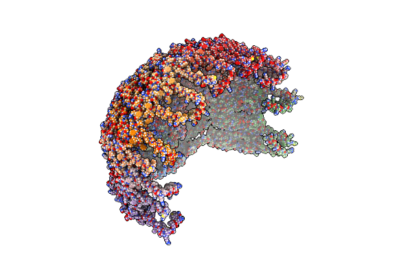
Organism: Puccinia graminis f. sp. tritici
Method: ELECTRON MICROSCOPY Release Date: 2025-09-24 Classification: ANTIFUNGAL PROTEIN |
 |
Organism: Homo sapiens
Method: ELECTRON MICROSCOPY Release Date: 2025-05-07 Classification: MEMBRANE PROTEIN |
 |
Organism: Homo sapiens
Method: ELECTRON MICROSCOPY Release Date: 2025-05-07 Classification: MEMBRANE PROTEIN |
 |
Organism: Agrobacterium fabrum str. c58
Method: ELECTRON MICROSCOPY Release Date: 2025-04-16 Classification: DNA BINDING PROTEIN |
 |
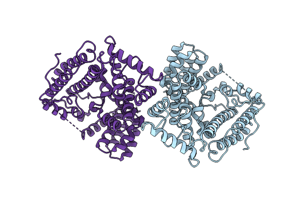
Organism: Streptomyces griseoviridis
Method: X-RAY DIFFRACTION Resolution:1.47 Å Release Date: 2025-02-19 Classification: TRANSFERASE Ligands: NCO, PGE, GOL, IPA, SAH, CL |
 |
Organism: Hordeum vulgare, Blumeria graminis
Method: ELECTRON MICROSCOPY Release Date: 2025-02-12 Classification: ANTIFUNGAL PROTEIN |
 |
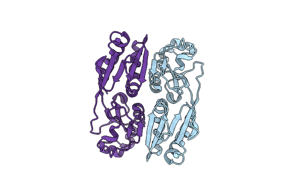
Organism: Rhodopirellula
Method: X-RAY DIFFRACTION Resolution:1.57 Å Release Date: 2025-01-08 Classification: HYDROLASE Ligands: MPO, SO4, CA |
 |
Crystal Structure Of Pbfuca From Planctomycetes Bacterium K23_9 In P 1 21 1
Organism: Planctomycetes bacterium k23_9
Method: X-RAY DIFFRACTION Resolution:2.50 Å Release Date: 2025-01-01 Classification: HYDROLASE Ligands: EDO |
 |
Crystal Structure Of Pbfuca From Planctomycetes Bacterium K23_9 In P 21 21 21
Organism: Planctomycetes bacterium k23_9
Method: X-RAY DIFFRACTION Resolution:1.40 Å Release Date: 2025-01-01 Classification: HYDROLASE Ligands: EDO |
 |
Crystal Structure Of Pbfuca From Planctomycetes Bacterium K23_9 In P 4 21 2
Organism: Planctomycetes bacterium k23_9
Method: X-RAY DIFFRACTION Resolution:2.18 Å Release Date: 2025-01-01 Classification: HYDROLASE Ligands: EDO, GOL |
 |
Organism: Clostridium beijerinckii
Method: ELECTRON MICROSCOPY Release Date: 2025-01-01 Classification: OXIDOREDUCTASE Ligands: ZN, SF4, 402 |
 |
Crystal Structure Of Apo-[Fefe]-Hydrogenase Cba5H From Clostridium Beijerinckii
Organism: Clostridium beijerinckii
Method: X-RAY DIFFRACTION Resolution:2.45 Å Release Date: 2024-12-18 Classification: OXIDOREDUCTASE Ligands: SF4, ZN, CL |
 |
Crystal Structure Of [Fefe]-Hydrogenase Cba5H From Clostridium Beijerinckii In Hinact State
Organism: Clostridium beijerinckii
Method: X-RAY DIFFRACTION Resolution:2.90 Å Release Date: 2024-12-18 Classification: OXIDOREDUCTASE Ligands: 402, SF4, ZN, CL |
 |
Organism: Homo sapiens
Method: X-RAY DIFFRACTION Resolution:2.38 Å Release Date: 2024-11-27 Classification: HYDROLASE Ligands: NAG, A1IJV, ZN |
 |
Organism: Homo sapiens
Method: X-RAY DIFFRACTION Resolution:1.99 Å Release Date: 2024-11-27 Classification: HYDROLASE Ligands: NAG, A1IJO, ZN |
 |
Organism: Homo sapiens
Method: X-RAY DIFFRACTION Resolution:2.20 Å Release Date: 2024-11-27 Classification: HYDROLASE Ligands: NAG, A1IJ3, ZN |
 |
Organism: Homo sapiens
Method: X-RAY DIFFRACTION Resolution:1.79 Å Release Date: 2024-11-27 Classification: HYDROLASE Ligands: A1IJ0, ZN |
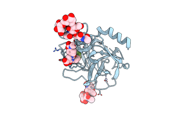 |
Crystal Structure Of Human Chymase In Complex With Fulacimstat (Compound86)
Organism: Homo sapiens
Method: X-RAY DIFFRACTION Resolution:1.80 Å Release Date: 2024-11-27 Classification: HYDROLASE Ligands: A1IJ2, ZN |