
Search Count: 18
 |
Nmr Solution Structure Of Subunit Epsilon Of The Mycobacterium Tuberculosis F-Atp Synthase
Organism: Mycobacterium tuberculosis (strain atcc 25618 / h37rv)
Method: SOLUTION NMR Release Date: 2018-10-10 Classification: PROTON TRANSPORT |
 |
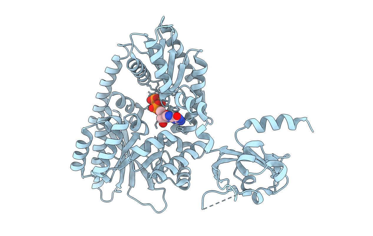
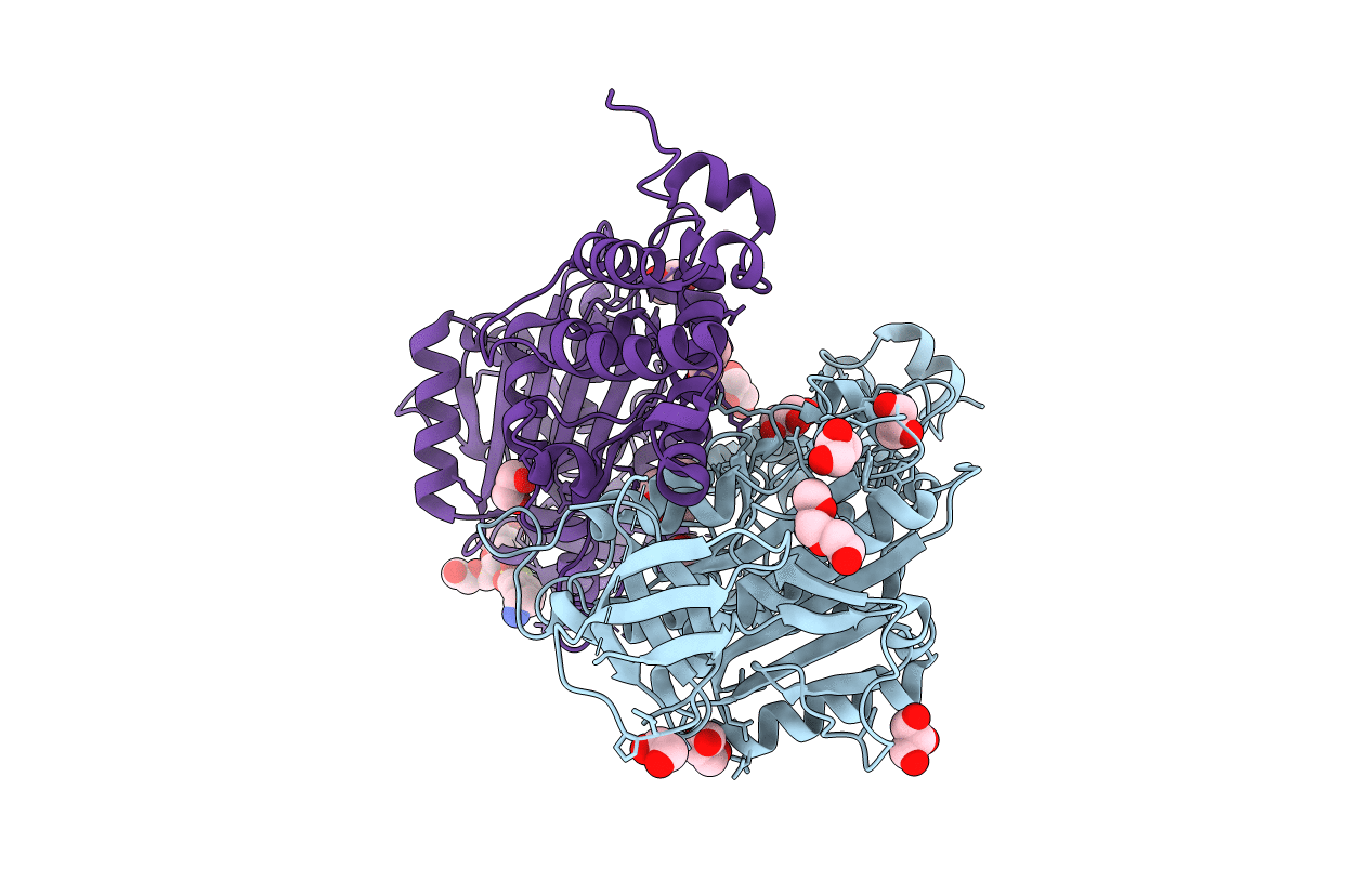
Crystal Structure Of The Pas-Ggdef-Eal Domain Of Pa0861 From Pseudomonas Aeruginosa
Organism: Pseudomonas aeruginosa (strain atcc 15692 / dsm 22644 / cip 104116 / jcm 14847 / lmg 12228 / 1c / prs 101 / pao1)
Method: X-RAY DIFFRACTION Resolution:2.28 Å Release Date: 2017-12-20 Classification: TRANSCRIPTION |
 |
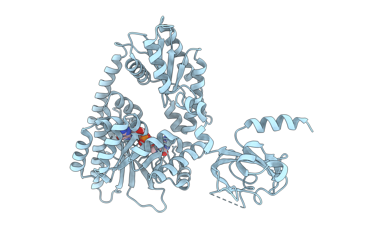
Crystal Structure Of The Pas-Ggdef-Eal Domain Of Pa0861 From Pseudomonas Aeruginosa In Complex With Gtp
Organism: Pseudomonas aeruginosa (strain atcc 15692 / dsm 22644 / cip 104116 / jcm 14847 / lmg 12228 / 1c / prs 101 / pao1)
Method: X-RAY DIFFRACTION Resolution:2.80 Å Release Date: 2017-12-20 Classification: TRANSCRIPTION Ligands: GTP, MG |
 |
Crystal Structure Of The Pas-Ggdef-Eal Domain Of Pa0861 From Pseudomonas Aeruginosa In Complex With Cyclic Di-Gmp
Organism: Pseudomonas aeruginosa (strain atcc 15692 / dsm 22644 / cip 104116 / jcm 14847 / lmg 12228 / 1c / prs 101 / pao1)
Method: X-RAY DIFFRACTION Resolution:3.31 Å Release Date: 2017-12-20 Classification: TRANSCRIPTION Ligands: C2E |
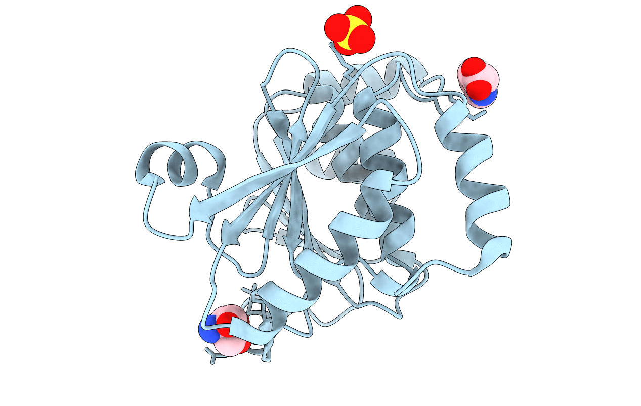 |
Crystal Structure Of The Ntd_N/C Domain Of Alkylhydroperoxide Reductase Ahpf From Enterococcus Faecalis (V583)
Organism: Enterococcus faecalis
Method: X-RAY DIFFRACTION Resolution:2.30 Å Release Date: 2017-11-22 Classification: OXIDOREDUCTASE Ligands: SO4, TRS, PRO |
 |
Crystallographic Structure Of The Enzymatically Active N-Terminal Domain Of The Rel Protein From Mycobacterium Tuberculosis
Organism: Mycobacterium tuberculosis (strain atcc 25618 / h37rv)
Method: X-RAY DIFFRACTION Resolution:3.70 Å Release Date: 2017-07-19 Classification: HYDROLASE, TRANSFERASE Ligands: MG |
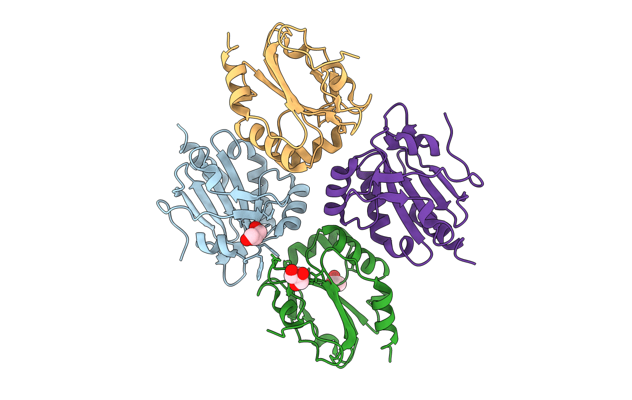 |
Asymmetry In The Active Site Of Mycobacterium Tuberculosis Ahpe Upon Exposure To Mycothiol
Organism: Mycobacterium tuberculosis h37rv
Method: X-RAY DIFFRACTION Resolution:2.43 Å Release Date: 2016-08-03 Classification: OXIDOREDUCTASE Ligands: GOL, ACT |
 |
Crystal Structure Of The Alkylhydroperoxide Reductase Subunit F (Ahpf) With Nadh From Escherichia Coli
Organism: Escherichia coli k12
Method: X-RAY DIFFRACTION Resolution:2.50 Å Release Date: 2015-07-15 Classification: OXIDOREDUCTASE Ligands: FAD, NAI, CD, SO4, GOL, PEG |
 |
Crystal Structure Of The Alkylhydroperoxide Reductase Subunit F (Ahpf) With Nad+ From Escherichia Coli
Organism: Escherichia coli k12
Method: X-RAY DIFFRACTION Resolution:2.40 Å Release Date: 2015-07-15 Classification: OXIDOREDUCTASE Ligands: FAD, NAD, SO4, CD, GOL, PEG |
 |
Organism: Saccharomyces cerevisiae
Method: X-RAY DIFFRACTION Resolution:2.33 Å Release Date: 2013-03-20 Classification: HYDROLASE Ligands: TRS, GOL |
 |
Organism: Methanosarcina mazei
Method: X-RAY DIFFRACTION Resolution:1.70 Å Release Date: 2012-09-05 Classification: HYDROLASE Ligands: GOL, CL, 1PE, AES, PEG, PGE |
 |
Organism: Methanosarcina mazei
Method: X-RAY DIFFRACTION Resolution:1.75 Å Release Date: 2012-09-05 Classification: HYDROLASE Ligands: GOL, P33, CL, AES, PG4, PG0 |
 |
Organism: Methanosarcina mazei
Method: X-RAY DIFFRACTION Resolution:1.75 Å Release Date: 2012-09-05 Classification: HYDROLASE Ligands: GOL, PG0, PG4, AES, 1PE, CL |
 |
Crystal Structure Of The C-Terminal Part Of Subunit E (E101-206) From Methanocaldococcus Jannaschii Of A1Ao Atp Synthase
Organism: Methanocaldococcus jannaschii
Method: X-RAY DIFFRACTION Resolution:4.10 Å Release Date: 2010-07-07 Classification: HYDROLASE |
 |
Expression, Purification, Spectroscopical And Crystallographical Studies Of Segments Of The Nucleotide Binding Domain Of The Reticulocyte Binding Protein Py235 Of Plasmodium Yoelii
Organism: Plasmodium yoelii yoelii
Method: X-RAY DIFFRACTION Resolution:4.00 Å Release Date: 2010-02-23 Classification: NUCLEOTIDE BINDING PROTEIN |
 |
A Second Transient Position Of Atp On Its Trail To The Nucleotide-Binding Site Of Subunit B Of The Motor Protein A1Ao Atp Synthase
Organism: Methanosarcina mazei
Method: X-RAY DIFFRACTION Resolution:3.43 Å Release Date: 2009-02-10 Classification: HYDROLASE Ligands: ATP, AES |
 |
Intermediate Position Of Atp On Its Trail To The Binding Pocket Inside The Subunit B Mutant R416W Of The Energy Converter A1Ao Atp Synthase
Organism: Methanosarcina mazei
Method: X-RAY DIFFRACTION Resolution:2.81 Å Release Date: 2008-09-09 Classification: HYDROLASE |
 |
Intermediate Position Of Atp On Its Trail To The Binding Pocket Inside The Subunit B Mutant R416W Of The Energy Converter A1Ao Atp Synthase
Organism: Methanosarcina mazei
Method: X-RAY DIFFRACTION Resolution:2.10 Å Release Date: 2008-09-09 Classification: HYDROLASE Ligands: ATP, AES, CIT |