
Search Count: 182
 |
Organism: Homo sapiens
Method: ELECTRON MICROSCOPY Release Date: 2025-12-17 Classification: IMMUNE SYSTEM |
 |
Organism: Sars-cov-2 pseudovirus, Homo sapiens
Method: ELECTRON MICROSCOPY Release Date: 2025-10-29 Classification: MEMBRANE PROTEIN/IMMUNE SYSTEM/INHIBITOR Ligands: A1AE8 |
 |
Organism: Severe acute respiratory syndrome coronavirus, Homo sapiens
Method: ELECTRON MICROSCOPY Release Date: 2025-10-29 Classification: MEMBRANE PROTEIN/IMMUNE SYSTEM/INHIBITOR |
 |
Organism: Middle east respiratory syndrome-related coronavirus, Homo sapiens
Method: ELECTRON MICROSCOPY Release Date: 2025-10-29 Classification: MEMBRANE PROTEIN/IMMUNE SYSTEM/INHIBITOR |
 |
Organism: Homo sapiens
Method: ELECTRON MICROSCOPY Release Date: 2025-10-22 Classification: IMMUNE SYSTEM Ligands: NAG |
 |
Organism: Homo sapiens
Method: ELECTRON MICROSCOPY Release Date: 2025-10-22 Classification: IMMUNE SYSTEM Ligands: NAG |
 |
Organism: Homo sapiens
Method: ELECTRON MICROSCOPY Release Date: 2025-10-22 Classification: IMMUNE SYSTEM |
 |
Organism: Homo sapiens
Method: ELECTRON MICROSCOPY Release Date: 2025-10-22 Classification: IMMUNE SYSTEM |
 |
Organism: Sfts virus hb29, Homo sapiens
Method: X-RAY DIFFRACTION Release Date: 2025-10-22 Classification: VIRAL PROTEIN Ligands: NAG |
 |
Organism: Homo sapiens
Method: ELECTRON MICROSCOPY Release Date: 2025-09-17 Classification: MEMBRANE PROTEIN Ligands: A1ETD |
 |
Organism: Homo sapiens
Method: ELECTRON MICROSCOPY Release Date: 2025-09-17 Classification: MEMBRANE PROTEIN Ligands: YN9, CLR, K |
 |
Organism: Homo sapiens
Method: ELECTRON MICROSCOPY Release Date: 2025-09-17 Classification: MEMBRANE PROTEIN Ligands: YN9, CLR, A1ETE, K |
 |
Organism: Hepatitis e virus (strain pakistan), Vicugna pacos
Method: X-RAY DIFFRACTION Release Date: 2025-09-03 Classification: VIRAL PROTEIN/IMMUNE SYSTEM |
 |
Organism: Horseshoe bat sarbecovirus
Method: ELECTRON MICROSCOPY Release Date: 2025-07-23 Classification: VIRAL PROTEIN Ligands: NAG, EIC |
 |
Organism: Coronaviridae
Method: ELECTRON MICROSCOPY Release Date: 2025-07-16 Classification: VIRAL PROTEIN Ligands: NAG |
 |
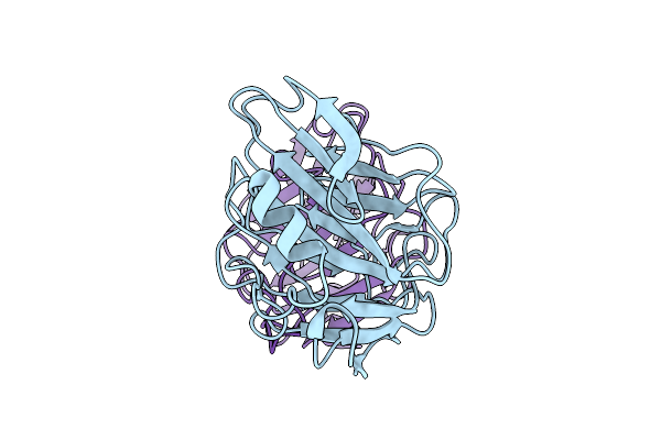
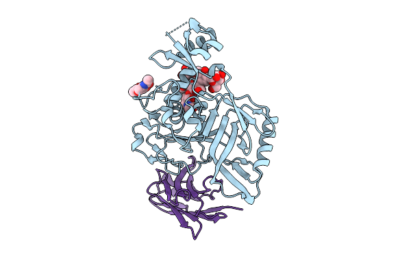
Molecular Basis Of Pathogenicity Of The Recently Emerged Fcov-23 Coronavirus. Complex Of Fapn With Fcov-23 Rbd
Organism: Felis catus, Feline coronavirus
Method: ELECTRON MICROSCOPY Release Date: 2025-07-09 Classification: VIRAL PROTEIN/HYDROLASE Ligands: NAG, ZN |
 |
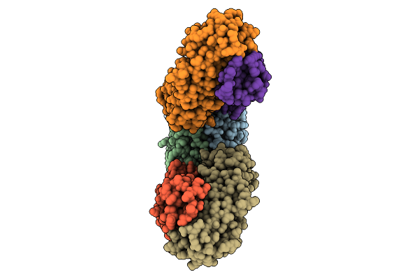
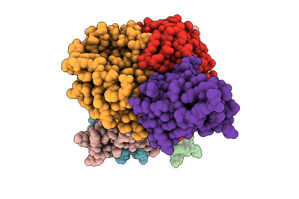
Molecular Basis Of Pathogenicity Of The Recently Emerged Fcov-23 Coronavirus. Fcov-23 S Short
Organism: Feline coronavirus
Method: ELECTRON MICROSCOPY Release Date: 2025-07-09 Classification: VIRAL PROTEIN Ligands: NAG, PAM |
 |
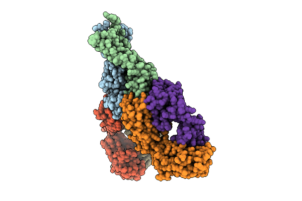
Molecular Basis Of Pathogenicity Of The Recently Emerged Fcov-23 Coronavirus. Fcov-23 S Do In Proximal Conformation (Local Refinement)
Organism: Feline coronavirus
Method: ELECTRON MICROSCOPY Release Date: 2025-07-09 Classification: VIRAL PROTEIN Ligands: NAG |
 |
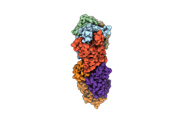
Molecular Basis Of Pathogenicity Of The Recently Emerged Fcov-23 Coronavirus. Fcov-23 S Long With Do In Swung-Out Conformation
Organism: Feline coronavirus
Method: ELECTRON MICROSCOPY Release Date: 2025-07-09 Classification: VIRAL PROTEIN Ligands: NAG, PAM |
 |
Molecular Basis Of Pathogenicity Of The Recently Emerged Fcov-23 Coronavirus. Fcov-23 S Long Domain 0 In Swung-Out Conformation (Local Refinement)
Organism: Feline coronavirus
Method: ELECTRON MICROSCOPY Release Date: 2025-07-09 Classification: VIRAL PROTEIN Ligands: NAG |