
Search Count: 32
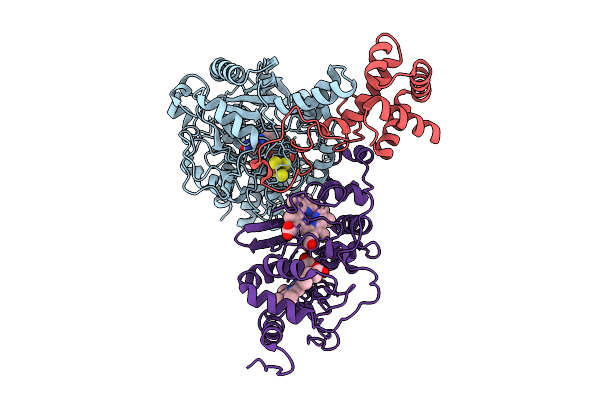 |
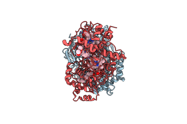
Cryo-Em Structure Of Fructose Dehydrogenase Variant From Gluconobacter Japonicus Truncating Heme 1C And C-Terminal Hydrophobic Regions
Organism: Gluconobacter japonicus
Method: ELECTRON MICROSCOPY Release Date: 2025-08-06 Classification: OXIDOREDUCTASE Ligands: FAD, F3S, HEC |
 |
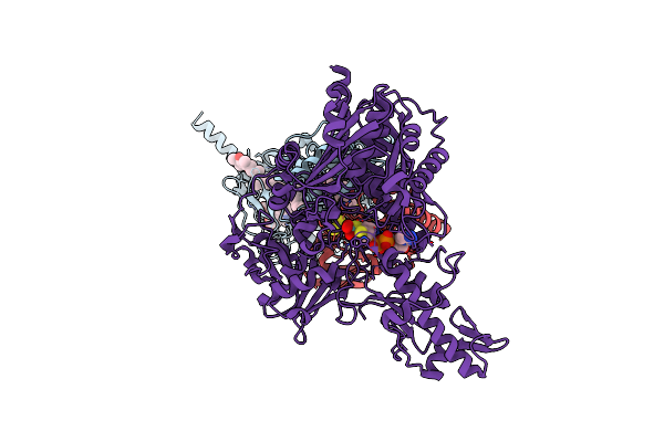
Structure Of The Salmonella Flagellar Flipqr Complex Reconstituted In The Peptidisc
Organism: Salmonella enterica subsp. enterica serovar typhimurium str. lt2
Method: ELECTRON MICROSCOPY Release Date: 2025-08-06 Classification: PROTEIN TRANSPORT |
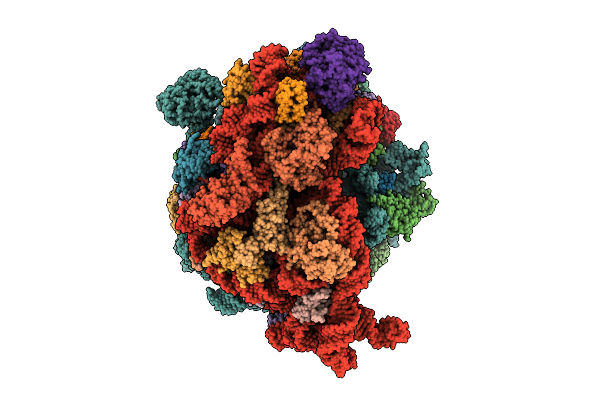 |
Cryo-Em Structure Of Escherichia Coli Hibernating Ribosome With Rnase I Mutant
Organism: Escherichia coli
Method: ELECTRON MICROSCOPY Release Date: 2025-07-30 Classification: RIBOSOME Ligands: MG, CA |
 |
Organism: Homo sapiens
Method: ELECTRON MICROSCOPY Release Date: 2025-03-26 Classification: IMMUNE SYSTEM |
 |
Single-Particle Cryo-Em Of Mycoplasma Pneumoniae Adhesin P1 Complexed With The Anti-Adhesive Fab Fragment.
Organism: Mycoplasmoides pneumoniae m129, Mus musculus
Method: ELECTRON MICROSCOPY Release Date: 2025-03-05 Classification: CELL ADHESION |
 |
Organism: Salmonella enterica subsp. enterica serovar typhimurium
Method: ELECTRON MICROSCOPY Release Date: 2025-01-01 Classification: MOTOR PROTEIN |
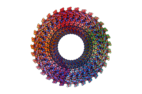 |
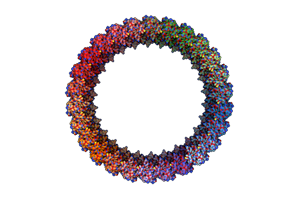
Structure Of The Rbm3 Ring Of Salmonella Flagellar Ms-Ring Protein Flif With C33 Symmetry Applied
Organism: Salmonella enterica subsp. enterica serovar typhimurium
Method: ELECTRON MICROSCOPY Release Date: 2025-01-01 Classification: MOTOR PROTEIN |
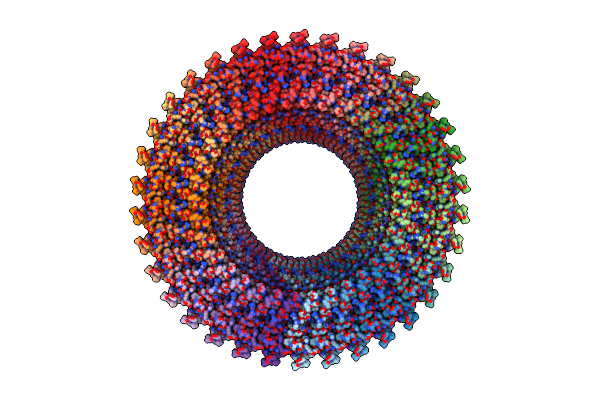 |
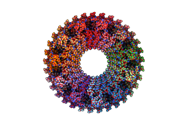
Structure Of The Rbm3 Ring Of Salmonella Flagellar Ms-Ring Protein Flif With C34 Symmetry Applied
Organism: Salmonella enterica subsp. enterica serovar typhimurium
Method: ELECTRON MICROSCOPY Release Date: 2025-01-01 Classification: MOTOR PROTEIN |
 |
Organism: Homo sapiens
Method: ELECTRON MICROSCOPY Release Date: 2024-09-04 Classification: IMMUNE SYSTEM |
 |
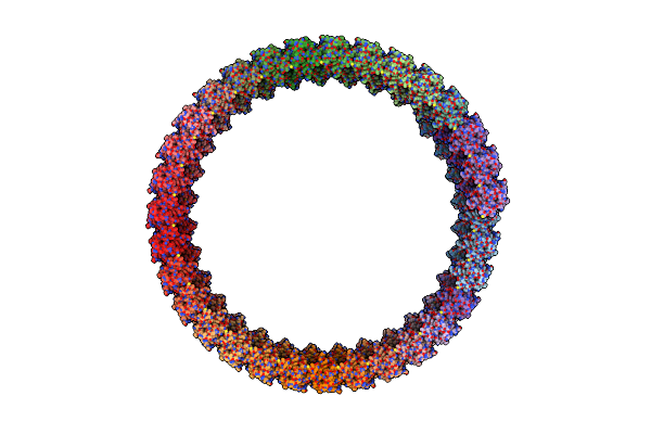
Salmonella Enterica Serovar Typhimurium Flic(G426A)Delta(204-292) Forming The L-Type Straight Filament
Organism: Salmonella enterica subsp. enterica serovar typhimurium str. lt2
Method: ELECTRON MICROSCOPY Release Date: 2024-08-07 Classification: MOTOR PROTEIN |
 |
Cryo-Em Structure Of Membrane-Bound Fructose Dehydrogenase From Gluconobacter Japonicus Variant-H1147A
Organism: Gluconobacter japonicus
Method: ELECTRON MICROSCOPY Release Date: 2024-05-22 Classification: OXIDOREDUCTASE Ligands: FAD, F3S, HEC, U10 |
 |
Cryo-Em Structure Of Membrane-Bound Fructose Dehydrogenase From Gluconobacter Japonicus Variant-N1146A
Organism: Gluconobacter japonicus
Method: ELECTRON MICROSCOPY Release Date: 2024-05-22 Classification: OXIDOREDUCTASE Ligands: FAD, F3S, HEC, U10 |
 |
Cryo-Em Structure Of Membrane-Bound Fructose Dehydrogenase From Gluconobacter Japonicus Variant-N1146Q
Organism: Gluconobacter japonicus
Method: ELECTRON MICROSCOPY Release Date: 2024-05-22 Classification: OXIDOREDUCTASE Ligands: FAD, F3S, HEC, U10 |
 |
Cryo-Em Structure Of Membrane-Bound Fructose Dehydrogenase From Gluconobacter Japonicus Variant-N1190A
Organism: Gluconobacter japonicus
Method: ELECTRON MICROSCOPY Release Date: 2024-05-22 Classification: OXIDOREDUCTASE Ligands: FAD, F3S, HEC, U10 |
 |
Cryo-Em Structure Of Na-Dithionite Reduced Membrane-Bound Fructose Dehydrogenase From Gluconobacter Japonicus
Organism: Gluconobacter japonicus
Method: ELECTRON MICROSCOPY Release Date: 2023-10-25 Classification: OXIDOREDUCTASE Ligands: FAD, F3S, HEC, U10 |
 |
Cryo-Em Structure Of K-Ferricyanide Oxidized Membrane-Bound Fructose Dehydrogenase From Gluconobacter Japonicus
Organism: Gluconobacter japonicus
Method: ELECTRON MICROSCOPY Release Date: 2023-10-25 Classification: OXIDOREDUCTASE Ligands: FAD, F3S, HEC, U10 |
 |
Cryo-Em Structure Of Membrane-Bound Alcohol Dehydrogenase From Gluconobacter Oxydans
Organism: Gluconobacter oxydans 621h, Gluconobacter oxydans
Method: ELECTRON MICROSCOPY Release Date: 2023-08-02 Classification: OXIDOREDUCTASE Ligands: HEC, PQQ, CA, U10 |
 |
Cryo-Em Structure Of Membrane-Bound Aldehyde Dehydrogenase From Gluconobacter Oxydans
Organism: Gluconobacter oxydans
Method: ELECTRON MICROSCOPY Release Date: 2023-08-02 Classification: OXIDOREDUCTASE Ligands: HEC, U10, FES, PCD |
 |
Cryo-Em Structure Of Membrane-Bound Fructose Dehydrogenase From Gluconobacter Japonicus
Organism: Gluconobacter japonicus
Method: ELECTRON MICROSCOPY Release Date: 2023-02-08 Classification: OXIDOREDUCTASE Ligands: FAD, F3S, HEC |
 |
Cryo-Em Structure Of Membrane-Bound Fructose Dehydrogenase From Gluconobacter Japonicus
Organism: Gluconobacter japonicus
Method: ELECTRON MICROSCOPY Release Date: 2022-11-30 Classification: OXIDOREDUCTASE Ligands: FAD, F3S, HEC |