
Search Count: 9
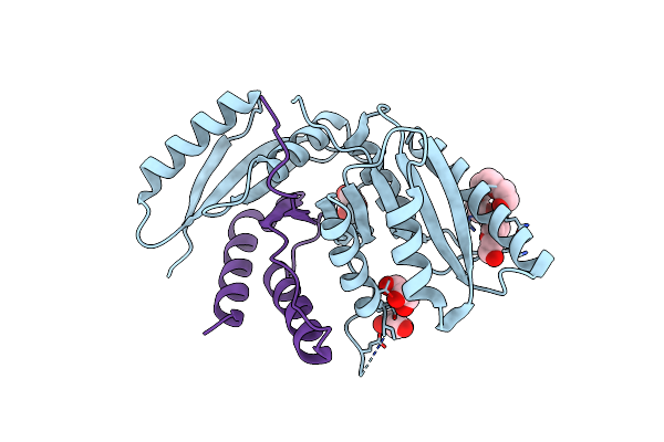 |
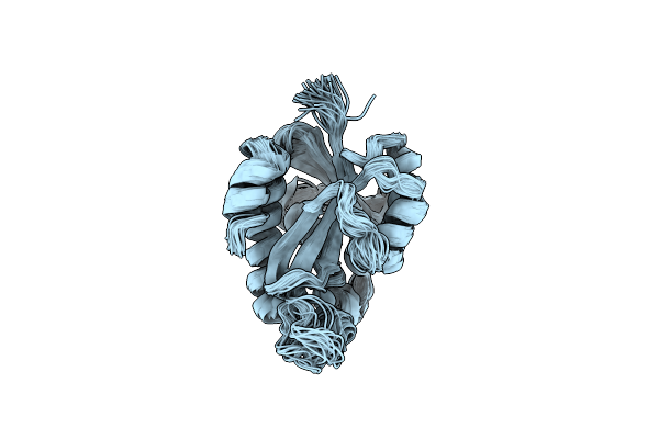
C-Terminal Regulatory Domain Of The Chloride Transporter Kcc-1 From C. Elegans, Proteolyzed During Crystallization
Organism: Caenorhabditis elegans
Method: X-RAY DIFFRACTION Resolution:1.80 Å Release Date: 2020-07-15 Classification: TRANSPORT PROTEIN Ligands: P33, GOL |
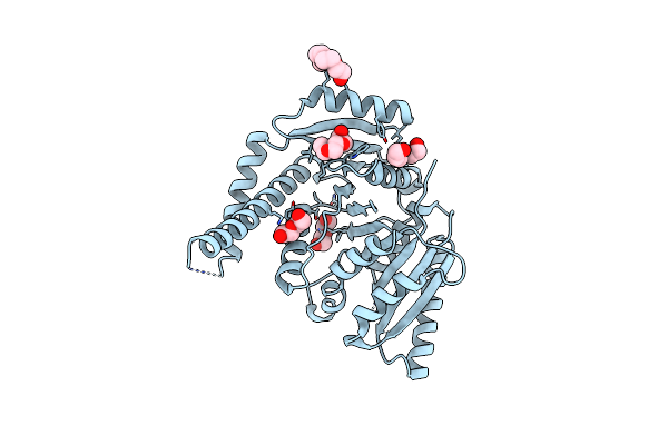 |
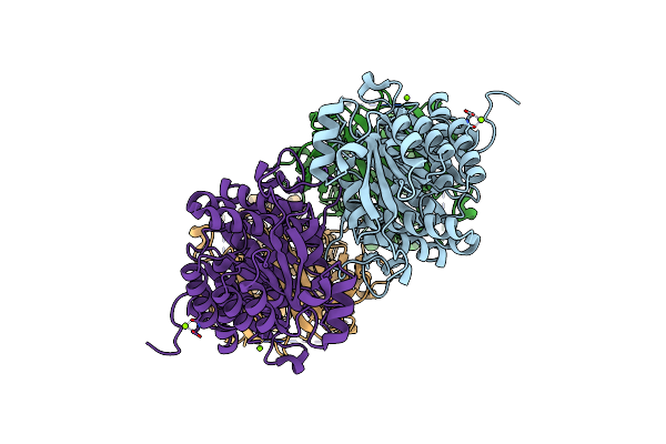
C-Terminal Regulatory Domain Of The Chloride Transporter Kcc-1 From C. Elegans
Organism: Caenorhabditis elegans
Method: X-RAY DIFFRACTION Resolution:2.20 Å Release Date: 2020-07-15 Classification: TRANSPORT PROTEIN Ligands: PGE, PEG, PG4 |
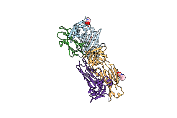 |
Organism: Homo sapiens
Method: X-RAY DIFFRACTION Resolution:2.38 Å Release Date: 2019-12-25 Classification: IMMUNE SYSTEM Ligands: NHE |
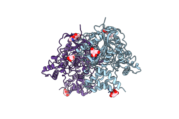 |
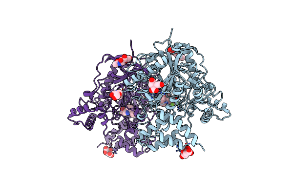
Crystal Structure Of An Insecticide-Resistant Acetylcholinesterase Mutant From The Malaria Vector Anopheles Gambiae In The Ligand-Free State
Organism: Anopheles gambiae
Method: X-RAY DIFFRACTION Resolution:2.30 Å Release Date: 2018-01-10 Classification: Hydrolase/Hydrolase Inhibitor Ligands: NAG, FLC, CL |
 |
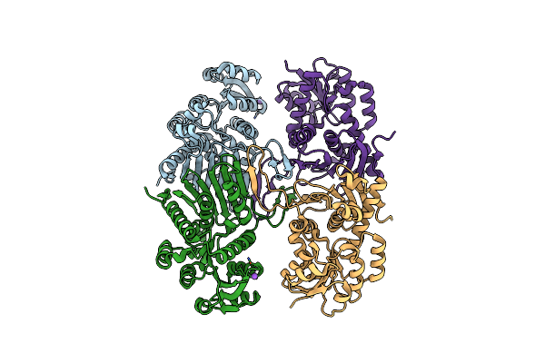
Crystal Structure Of An Insecticide-Resistant Acetylcholinesterase Mutant From The Malaria Vector Anopheles Gambiae In Complex With A Difluoromethyl Ketone Inhibitor
Organism: Anopheles gambiae
Method: X-RAY DIFFRACTION Resolution:2.26 Å Release Date: 2018-01-10 Classification: HYDROLASE/Hydrolase Inhibitor Ligands: BT7, NAG, FLC, CL |
 |
Crystal Structure Of A Periplasmic Sugar-Binding Protein From The Pseudomonas Fluorescens
Organism: Pseudomonas fluorescens pf-5
Method: X-RAY DIFFRACTION Resolution:1.60 Å Release Date: 2009-05-12 Classification: SUGAR BINDING PROTEIN Ligands: SO4, GOL |
 |
Crystal Structure Of A Myo-Inositol Dehydrogenase From Corynebacterium Glutamicum Atcc 13032
Organism: Corynebacterium glutamicum
Method: X-RAY DIFFRACTION Resolution:2.30 Å Release Date: 2008-10-21 Classification: OXIDOREDUCTASE |
 |
Organism: Pseudomonas syringae pv. tomato
Method: X-RAY DIFFRACTION Resolution:2.04 Å Release Date: 2008-08-05 Classification: OXIDOREDUCTASE Ligands: MG |
 |
Solution Structure Of Peptidyl-Trna Hydrolase From Mycobacterium Tuberculosis H37Rv.
Organism: Mycobacterium tuberculosis
Method: SOLUTION NMR Release Date: 2008-05-27 Classification: HYDROLASE |