
Search Count: 53
 |
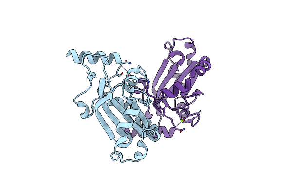
Organism: Homo sapiens
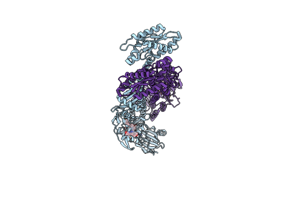
Method: X-RAY DIFFRACTION Release Date: 2024-03-06 Classification: TRANSPORT PROTEIN Ligands: PEG, TSS |
 |
Organism: Vibrio cholerae
Method: SOLUTION NMR Release Date: 2021-10-20 Classification: TRANSCRIPTION |
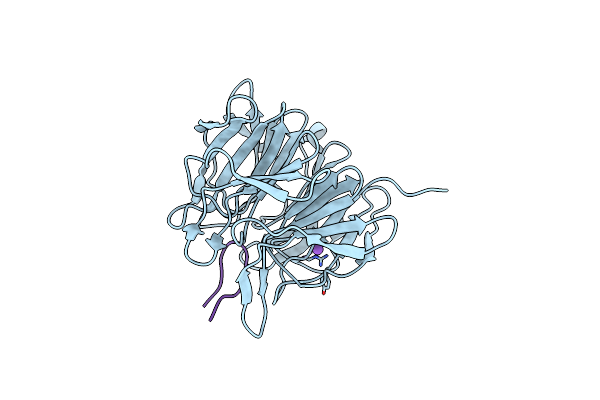 |
Crystal Structure Of The Kelch Domain In Complex With 11 Amino Acid Peptide (Model Of The Etge Loop)
Organism: Homo sapiens
Method: X-RAY DIFFRACTION Resolution:2.75 Å Release Date: 2020-09-16 Classification: LIGASE |
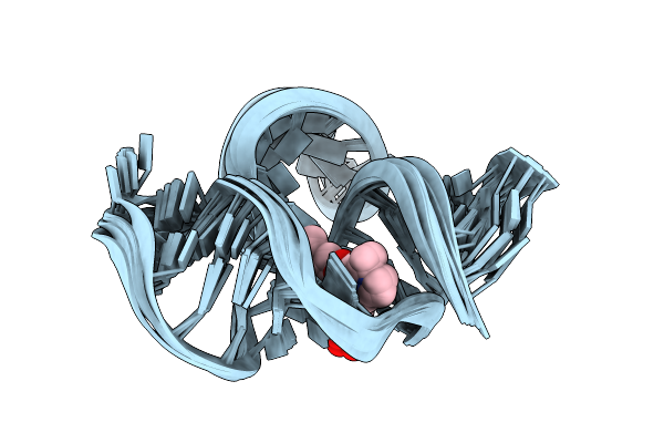 |
Solution Nmr Structure Of The Tetramethylrhodamine (Tmr) Aptamer 3 In Complex With 5-Tamra
Organism: Synthetic construct
Method: SOLUTION NMR Release Date: 2019-07-17 Classification: RNA Ligands: FH8 |
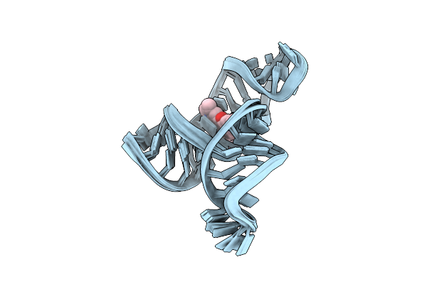 |
Solution Nmr Structure Of The Tetramethylrhodamine (Tmr) Aptamer 3 In Complex With 5-Tamra
Organism: Synthetic construct
Method: SOLUTION NMR Release Date: 2019-07-17 Classification: RNA Ligands: FH8 |
 |
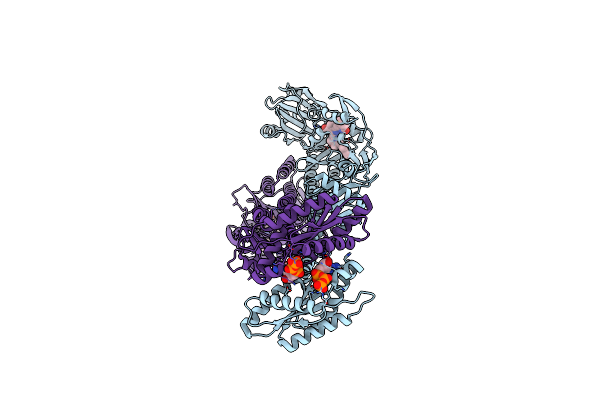
Organism: Saccharomyces cerevisiae
Method: X-RAY DIFFRACTION Resolution:1.55 Å Release Date: 2019-03-27 Classification: CHAPERONE Ligands: MG |
 |
Organism: Homo sapiens
Method: X-RAY DIFFRACTION Resolution:1.65 Å Release Date: 2019-03-27 Classification: CHAPERONE Ligands: PO4 |
 |
Organism: Saccharomyces cerevisiae
Method: X-RAY DIFFRACTION Resolution:2.80 Å Release Date: 2019-03-27 Classification: CHAPERONE Ligands: MG |
 |
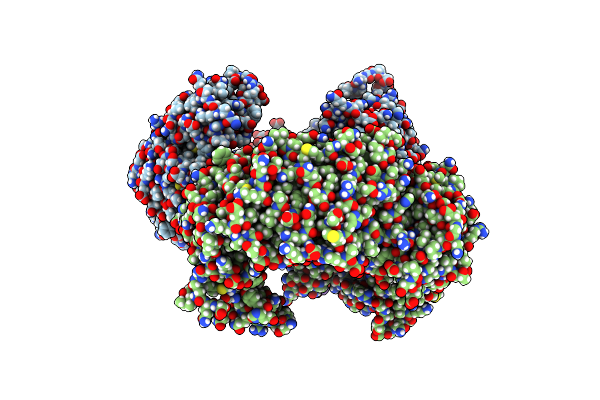
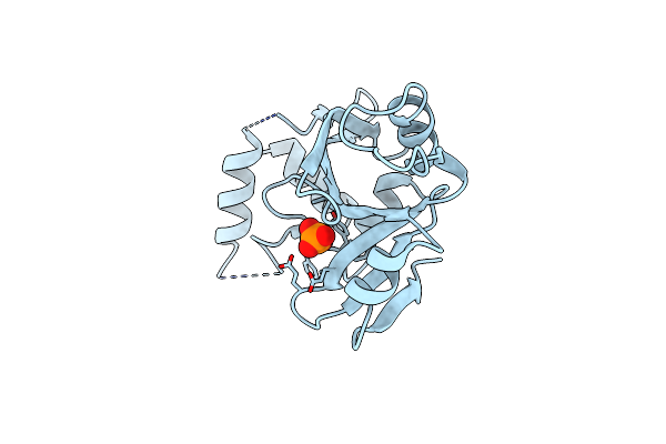
Structure Of Human Nad(P) H:Quinone Oxidoreductase In Complex With N-(2-Bromophenyl)Pyrrolidine-1-Sulfonamide
Organism: Homo sapiens
Method: X-RAY DIFFRACTION Resolution:2.76 Å Release Date: 2019-03-20 Classification: OXIDOREDUCTASE Ligands: FAD, EAW |
 |
Solution Structure Of Oxidized And Amidated Human Iapp (1-37), The Diabetes Ii Peptide.
|
 |
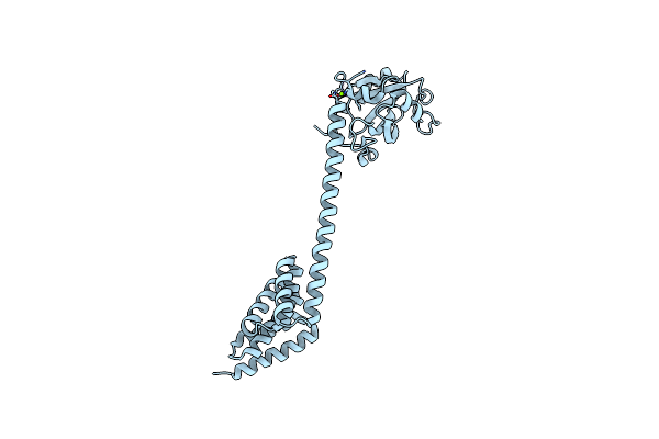
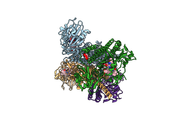
Bacteriophytochrome Activated Diguanylyl Cyclase From Idiomarina Species A28L
Organism: Idiomarina sp. a28l
Method: X-RAY DIFFRACTION Resolution:3.00 Å Release Date: 2017-03-15 Classification: TRANSFERASE Ligands: LBV, CL |
 |
Bacteriophytochrome Activated Diguanylyl Cyclase From Idiomarina Species A28L With Gtp Bound
Organism: Idiomarina sp. a28l
Method: X-RAY DIFFRACTION Resolution:2.80 Å Release Date: 2017-03-15 Classification: TRANSFERASE Ligands: LBV, CL, GTP, MG |
 |
Photosensory Module Of Bacteriophytochrome Linked Diguanylyl Cyclase From Idiomarina Species A28L
Organism: Idiomarina sp. a28l
Method: X-RAY DIFFRACTION Resolution:2.40 Å Release Date: 2017-03-15 Classification: SIGNALING PROTEIN Ligands: LBV, CL, GOL |
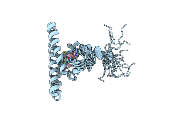 |
Organism: Homo sapiens
Method: SOLUTION NMR Release Date: 2017-03-15 Classification: CHAPERONE Ligands: FAR |
 |
Crystal Structure Of Mouse Phospholipid Hydroperoxide Glutathione Peroxidase 4 (Gpx4)
Organism: Mus musculus
Method: X-RAY DIFFRACTION Resolution:1.80 Å Release Date: 2016-10-19 Classification: OXIDOREDUCTASE Ligands: EDO |
 |
Crystal Structure Of The R139W Variant Of Human Nad(P)H:Quinone Oxidoreductase
Organism: Homo sapiens
Method: X-RAY DIFFRACTION Resolution:2.09 Å Release Date: 2016-06-29 Classification: OXIDOREDUCTASE Ligands: FAD, BTB |
 |
Organism: Drosophila melanogaster
Method: X-RAY DIFFRACTION Resolution:2.60 Å Release Date: 2016-01-20 Classification: DNA BINDING PROTEIN Ligands: CL |
 |
Crystal Structure Of D. Melanogaster Pur-Alpha Repeat I-Ii In Complex With Dna.
Organism: Drosophila melanogaster
Method: X-RAY DIFFRACTION Resolution:2.00 Å Release Date: 2016-01-20 Classification: DNA BINDING PROTEIN Ligands: CL, SO4 |
 |
Nmr Solution Structure Of The C-Terminal Domain Of Nisi, A Lipoprotein From Lactococcus Lactis Which Confers Immunity Against Nisin
Organism: Lactococcus lactis subsp. lactis
Method: SOLUTION NMR Release Date: 2015-10-21 Classification: LANTIBIOTIC-BINDING PROTEIN |
 |
Nmr Solution Structure Of The N-Terminal Domain Of Nisi, A Lipoprotein From Lactococcus Lactis Which Confers Immunity Against Nisin
Organism: Lactococcus lactis subsp. lactis
Method: SOLUTION NMR Release Date: 2015-10-21 Classification: LANTIBIOTIC-BINDING PROTEIN |