
Search Count: 1,401
 |
Organism: Arabidopsis thaliana
Method: X-RAY DIFFRACTION Release Date: 2025-12-24 Classification: CELL CYCLE Ligands: BEZ, NA |
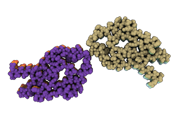 |
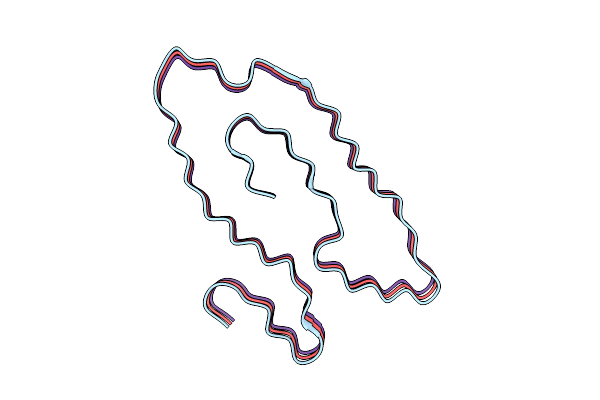
Cryo-Em Structure Of Renal Amyloid Fibril From An Immunoglobulin Light Chain Amyloidosis Patient In Polymorph B
Organism: Homo sapiens
Method: ELECTRON MICROSCOPY Release Date: 2025-12-24 Classification: PROTEIN FIBRIL |
 |
Cryo-Em Structure Of Renal Amyloid Fibril From An Immunoglobulin Light Chain Amyloidosis Patient In Polymorph A
Organism: Homo sapiens
Method: ELECTRON MICROSCOPY Release Date: 2025-12-24 Classification: PROTEIN FIBRIL |
 |
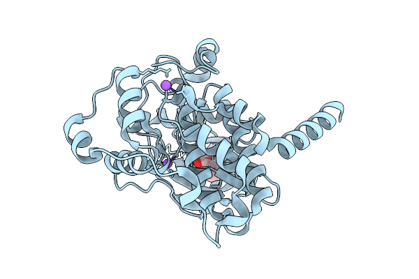
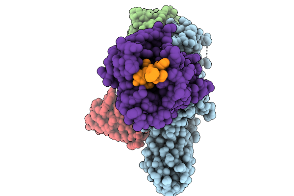
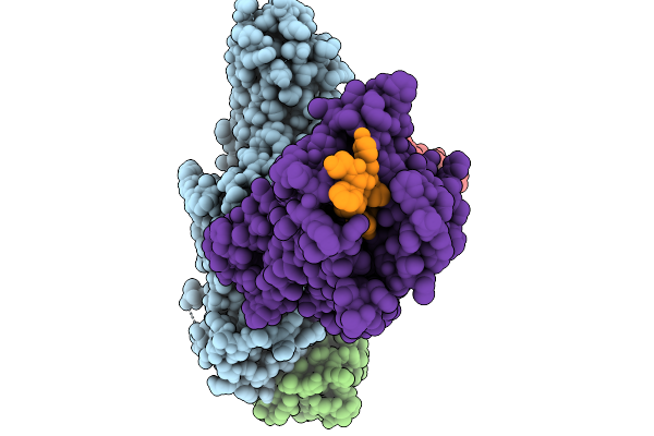
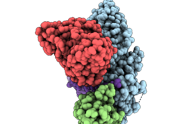
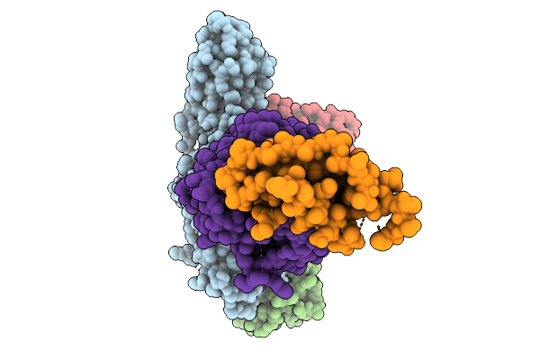
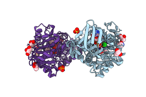
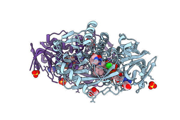
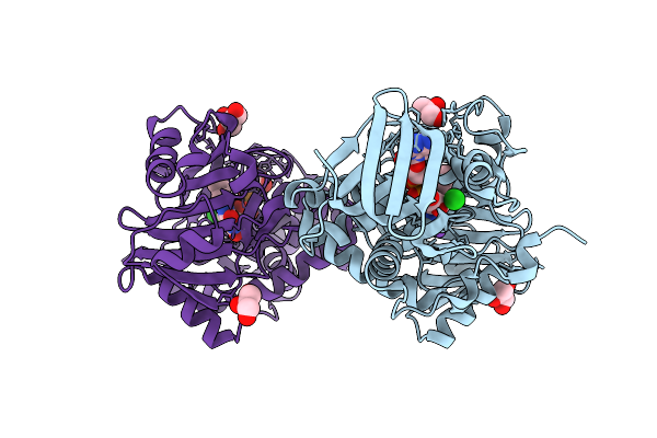
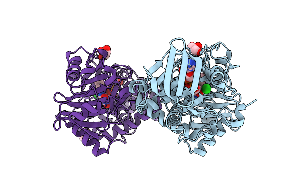
Arabidopsis Thaliana Casein Kinase 2 (Ck2) Alpha1 - Beta1 Complex Bound To Inositol Hexakisphosphate (Insp6)
Organism: Arabidopsis thaliana
Method: X-RAY DIFFRACTION Release Date: 2025-12-17 Classification: CELL CYCLE Ligands: IHP, ZN |
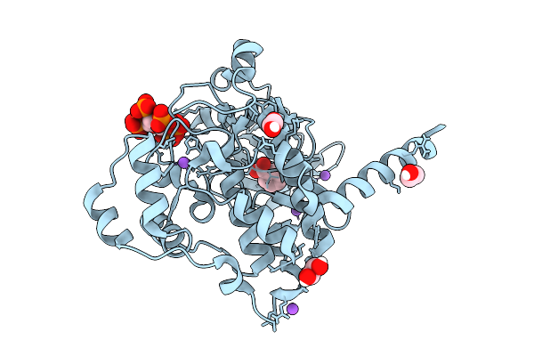 |
Arabidopsis Thaliana Casein Kinase 2 (Ck2) Alpha Isoform 1 In Complex With Inositol Hexakisphosphate (Insp6)
Organism: Arabidopsis thaliana
Method: X-RAY DIFFRACTION Release Date: 2025-12-10 Classification: CELL CYCLE Ligands: IHP, BEZ, EDO, CL, NA |
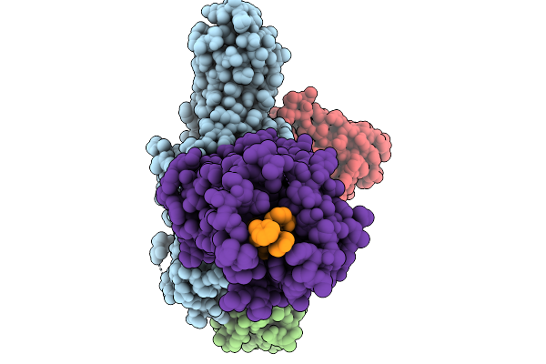 |
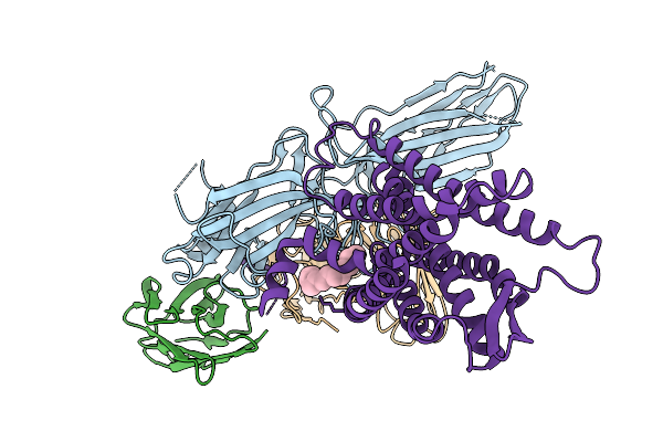
Cryo-Em Structure Of The G Protein-Coupled Receptor 1 (Gpr1) Bound To Chemerin And Beta-Arrestin 1 (Conformation 1)
Organism: Homo sapiens, Escherichia phage ecszw-2
Method: ELECTRON MICROSCOPY Release Date: 2025-11-19 Classification: MEMBRANE PROTEIN/IMMUNE SYSTEM |
 |
Cryo-Em Structure Of The G Protein-Coupled Receptor 1 (Gpr1) Bound To Chemerin And Beta-Arrestin 1 (Conformation 2)
Organism: Homo sapiens, Escherichia phage ecszw-2
Method: ELECTRON MICROSCOPY Release Date: 2025-11-19 Classification: MEMBRANE PROTEIN/IMMUNE SYSTEM |
 |
Cryo-Em Structure Of The G Protein-Coupled Receptor 1 (Gpr1) Bound To Chemerin And Beta-Arrestin 1 (Conformation 3)
Organism: Homo sapiens, Escherichia phage ecszw-2
Method: ELECTRON MICROSCOPY Release Date: 2025-11-19 Classification: MEMBRANE PROTEIN/IMMUNE SYSTEM |
 |
Cryo-Em Structure Of The G Protein-Coupled Receptor 1 (Gpr1) Bound To Chemerin And Beta-Arrestin 1 (Conformation 4)
Organism: Homo sapiens, Escherichia phage ecszw-2
Method: ELECTRON MICROSCOPY Release Date: 2025-11-19 Classification: MEMBRANE PROTEIN/IMMUNE SYSTEM |
 |
Composite Map Of The G Protein-Coupled Receptor 1 (Gpr1) Bound To Chemerin And Beta-Arrestin 2
Organism: Homo sapiens, Escherichia phage ecszw-2
Method: ELECTRON MICROSCOPY Release Date: 2025-11-19 Classification: MEMBRANE PROTEIN/IMMUNE SYSTEM Ligands: Y01 |
 |
Cryo-Em Structure Of The G Protein-Coupled Receptor 1 (Gpr1) Bound To Beta-Arrestin 1 In Ligand-Free State
Organism: Homo sapiens, Escherichia phage ecszw-2
Method: ELECTRON MICROSCOPY Release Date: 2025-11-19 Classification: MEMBRANE PROTEIN/IMMUNE SYSTEM Ligands: PAM |
 |
Organism: Homo sapiens
Method: ELECTRON MICROSCOPY Release Date: 2025-10-29 Classification: PROTEIN FIBRIL |
 |
Organism: Homo sapiens
Method: ELECTRON MICROSCOPY Release Date: 2025-10-29 Classification: PROTEIN FIBRIL |
 |
Organism: Euglena gracilis
Method: ELECTRON MICROSCOPY Release Date: 2025-10-08 Classification: PHOTOSYNTHESIS Ligands: CL0, CLA, PQN, SF4, BCR, LHG, UNL, DGD, DD6 |
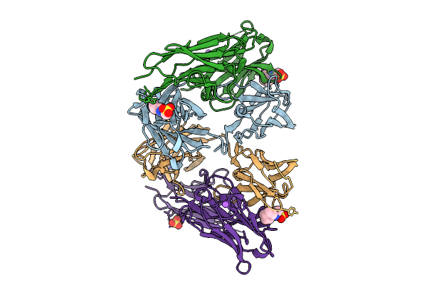 |
Crystal Structure Of A High Affinity Vl-Vh Tetrabody For The Erythropoietin Receptor
Organism: Homo sapiens
Method: X-RAY DIFFRACTION Release Date: 2025-10-01 Classification: SIGNALING PROTEIN Ligands: NHE, CL, SO4, NA |
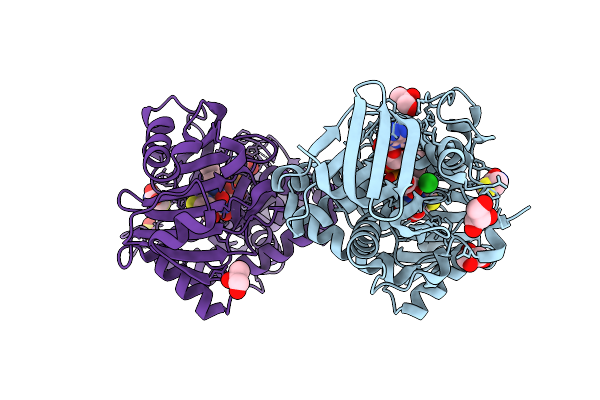 |
Monomeric Sarcosine Oxidase From Bacillus Sp. (Soxb) Complexed With L-Thioproline
Organism: Bacillus sp. b-0618
Method: X-RAY DIFFRACTION Release Date: 2025-09-24 Classification: OXIDOREDUCTASE Ligands: FAD, PRS, CL, GOL, SO4 |
 |
Monomeric Sarcosine Oxidase From Bacillus Sp. (Soxb) Complexed With L-Proline
Organism: Bacillus sp. b-0618
Method: X-RAY DIFFRACTION Release Date: 2025-09-24 Classification: OXIDOREDUCTASE Ligands: FAD, PRO, GOL, CL, SO4, PO4 |
 |
Monomeric Sarcosine Oxidase From Bacillus Sp. (Soxb) Complexed With D-Proline
Organism: Bacillus sp. b-0618
Method: X-RAY DIFFRACTION Release Date: 2025-09-24 Classification: OXIDOREDUCTASE Ligands: FAD, DPR, CL, GOL, SO4 |
 |
Organism: Bacillus sp. b-0618
Method: X-RAY DIFFRACTION Release Date: 2025-09-24 Classification: OXIDOREDUCTASE Ligands: FAD, GOL, CL, NA |
 |
Organism: Bacillus sp. b-0618
Method: X-RAY DIFFRACTION Release Date: 2025-09-24 Classification: OXIDOREDUCTASE Ligands: FAD, GOL, CL |