
Search Count: 205
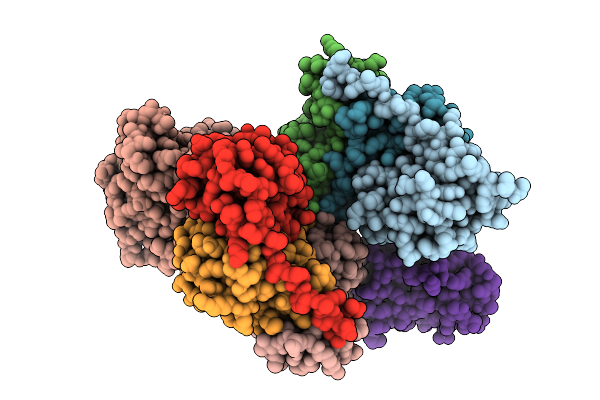 |
Crystal Structure Of Compound 3-Mediated Ternary Complex Of Kras G12V C118S Gdp With Pvhl:Elonginc:Elonginb
Organism: Homo sapiens
Method: X-RAY DIFFRACTION Release Date: 2025-11-12 Classification: ONCOPROTEIN Ligands: A1JH1, GDP, MG |
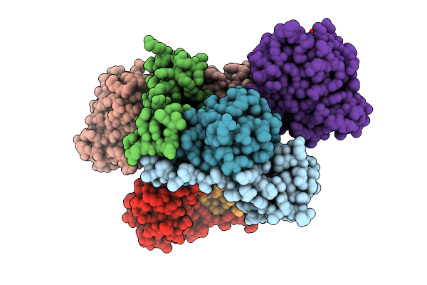 |
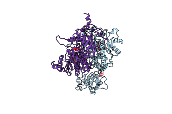
Crystal Structure Of Acbi4-Mediated Ternary Complex Of Kras G12D C118S Gdp With Pvhl:Elonginc:Elonginb
Organism: Homo sapiens
Method: X-RAY DIFFRACTION Release Date: 2025-11-12 Classification: ONCOPROTEIN Ligands: A1JHI, GDP, MG |
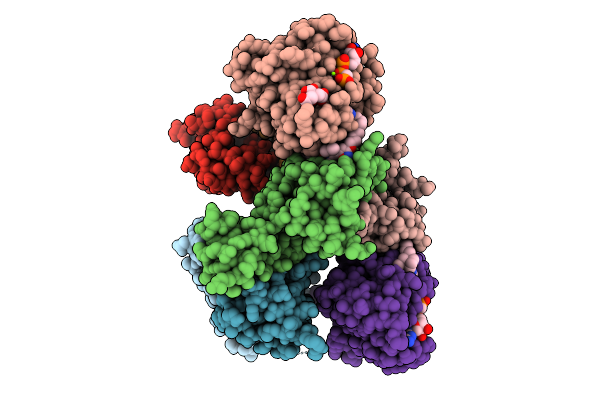 |
Crystal Structure Of Compound 1-Mediated Ternary Complex Of Kras G12R Gcp With Pvhl:Elonginc:Elonginb
Organism: Homo sapiens
Method: X-RAY DIFFRACTION Release Date: 2025-11-12 Classification: ONCOPROTEIN Ligands: GOL, X53, GCP, MG, FLC |
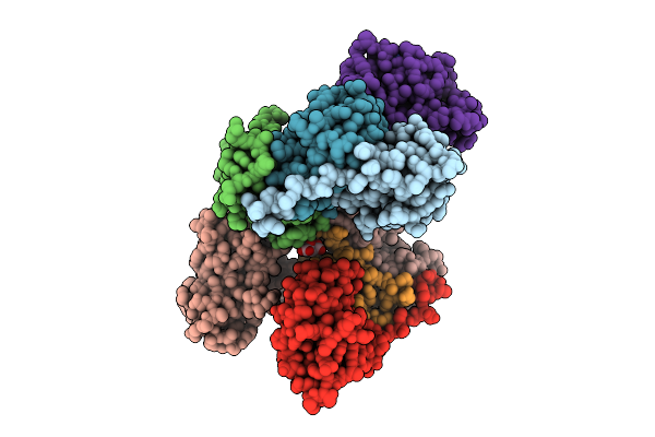 |
Crystal Structure Of Compound 3-Mediated Ternary Complex Of Kras G12R Gcp With Pvhl:Elonginc:Elonginb
Organism: Homo sapiens
Method: X-RAY DIFFRACTION Release Date: 2025-11-12 Classification: ONCOPROTEIN Ligands: FLC, A1JH1, GCP, MG, GOL |
 |
Crystal Structure Of Acbi4-Mediated Ternary Complex Of Kras G12R Gcp With Pvhl:Elonginc:Elonginb
Organism: Homo sapiens
Method: X-RAY DIFFRACTION Release Date: 2025-11-12 Classification: ONCOPROTEIN Ligands: SO4, A1JHI, GCP, MG |
 |
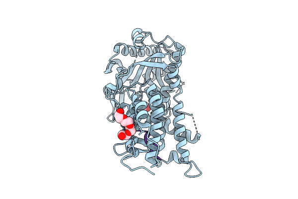
Crystal Structure Of Lysyl-Trna Synthetase From Plasmodium Falciparum Complexed With Lys-Ams
Organism: Plasmodium falciparum
Method: X-RAY DIFFRACTION Release Date: 2025-09-10 Classification: LIGASE Ligands: KAA |
 |
Organism: Thermobifida fusca yx
Method: X-RAY DIFFRACTION Release Date: 2025-08-06 Classification: HYDROLASE |
 |
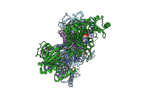
Organism: Candidatus odinarchaeota
Method: ELECTRON MICROSCOPY Release Date: 2025-07-30 Classification: CELL CYCLE Ligands: G2P |
 |
Organism: Thermobifida fusca yx
Method: X-RAY DIFFRACTION Release Date: 2025-07-23 Classification: HYDROLASE Ligands: CL, NA, GOL |
 |
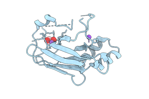
Influenza Polymerase A C-Terminal Domain Of Pa Subunit With Peptide Inhibitor Containing Two Norleucines
Organism: Influenza a virus (a/california/07/2009(h1n1)), Synthetic construct
Method: X-RAY DIFFRACTION Release Date: 2025-07-16 Classification: VIRAL PROTEIN Ligands: EDO, PG4 |
 |
Influenza Polymerase A C-Terminal Domain Of Pa Subunit With Stapled Peptide Inhibitor
Organism: Influenza a virus (a/california/07/2009(h1n1))
Method: X-RAY DIFFRACTION Release Date: 2025-07-16 Classification: VIRAL PROTEIN Ligands: PGE, EDO, A1IG4 |
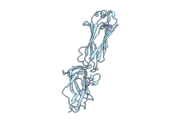 |
Crystal Structure Of C-Terminal Rigid Fragment Containing Middle And C-Terminal Domains Of The Basal Pilin Ebpb From Enterococcus Faecalis.
Organism: Enterococcus faecalis og1rf
Method: X-RAY DIFFRACTION Release Date: 2025-07-16 Classification: CELL ADHESION |
 |
Crystal Structure Of N-Terminal Flexible Domain Of The Shaft Pilin Ebpc From Enterococcus Faecalis.
Organism: Enterococcus faecalis og1rf
Method: X-RAY DIFFRACTION Release Date: 2025-07-16 Classification: CELL ADHESION |
 |
Crystal Structure Of N-Terminal Flexible Domain Of The Shaft Pilin Ebpc From Enterococcus Faecalis.
Organism: Enterococcus faecalis og1rf
Method: X-RAY DIFFRACTION Release Date: 2025-07-16 Classification: CELL ADHESION |
 |
Organism: Enterococcus faecalis og1rf
Method: X-RAY DIFFRACTION Release Date: 2025-07-16 Classification: CELL ADHESION |
 |
Crystal Structure Of C-Terminal Stable Fragment Of The Shaft Pilin Ebpc From Enterococcus Faecalis
Organism: Enterococcus faecalis og1rf
Method: X-RAY DIFFRACTION Release Date: 2025-07-16 Classification: CELL ADHESION Ligands: MG, NA |
 |
Crystal Structure Of The Basal Pilin Ebpb From Enterococcus Faecalis With A Partially Disordered N-Terminal Domain.
Organism: Enterococcus faecalis og1rf
Method: X-RAY DIFFRACTION Release Date: 2025-07-16 Classification: CELL ADHESION |
 |
Organism: Thermobifida fusca yx
Method: X-RAY DIFFRACTION Release Date: 2025-07-09 Classification: HYDROLASE Ligands: EDO, CL, NA |
 |
Organism: Plasmodium vivax
Method: X-RAY DIFFRACTION Release Date: 2025-06-04 Classification: LIGASE Ligands: SO4, EDO |
 |
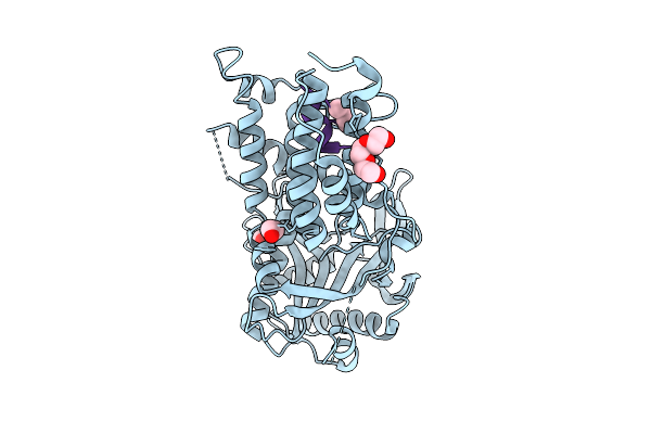
Plasmodium Vivax Aspartyl-Trna Synthetase In Complex With Aspartyl-Adenylate (Asp-Amp) Complex.
Organism: Plasmodium vivax
Method: X-RAY DIFFRACTION Release Date: 2025-06-04 Classification: LIGASE Ligands: AMO, GOL, ACT |