
Search Count: 4,561
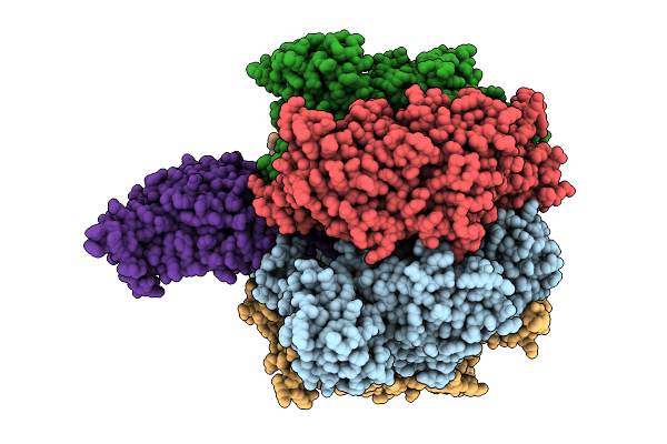 |
Organism: Bacillus sp. ps3
Method: ELECTRON MICROSCOPY Release Date: 2025-11-05 Classification: ELECTRON TRANSPORT Ligands: MG, ATP, PO4, ADP |
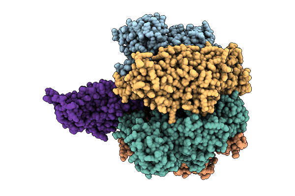 |
Organism: Bacillus sp. ps3, Pseudomonas aeruginosa
Method: ELECTRON MICROSCOPY Release Date: 2025-11-05 Classification: ELECTRON TRANSPORT Ligands: ATP, MG, ADP |
 |
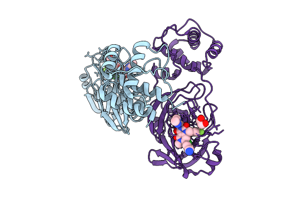
Crystal Structure Of Kirsten Rat Sarcoma G12C Complexed With Gmppnp And Covalently Bound To An Adduct Of {(2S)-4-[7-(8-Chloronaphthalen-1-Yl)-2-{[(2S)-1-Methylpyrrolidin-2-Yl]Methoxy}-5,6,7,8-Tetrahydropyrido[3,4-D]Pyrimidin-4-Yl]-1-[(2Z)-2-Fluoro-3-(Pyridin-2-Yl)Prop-2-Enoyl]Piperazin-2-Yl}Acetonitrile
Organism: Homo sapiens
Method: X-RAY DIFFRACTION Release Date: 2025-11-05 Classification: HYDROLASE/HYDROLASE INHIBITOR Ligands: GNP, A1B7P, MG |
 |
Crystal Structure Of Kirsten Rat Sarcoma G12C Complexed With Gdp And Covalently Bound To An Adduct Of (2S)-1-{4-[(7P)-7-(8-Ethynyl-7-Fluoro-3-Hydroxynaphthalen-1-Yl)-8-Fluoro-2-{[(2R,4R,7As)-2-Fluorotetrahydro-1H-Pyrrolizin-7A(5H)-Yl]Methoxy}Pyrido[4,3-D]Pyrimidin-4-Yl]Piperazin-1-Yl}-2-Fluoro-3-(1,3-Thiazol-2-Yl)Propan-1-One
Organism: Homo sapiens
Method: X-RAY DIFFRACTION Release Date: 2025-11-05 Classification: HYDROLASE/HYDROLASE INHIBITOR Ligands: GDP, A1B7Q, MG |
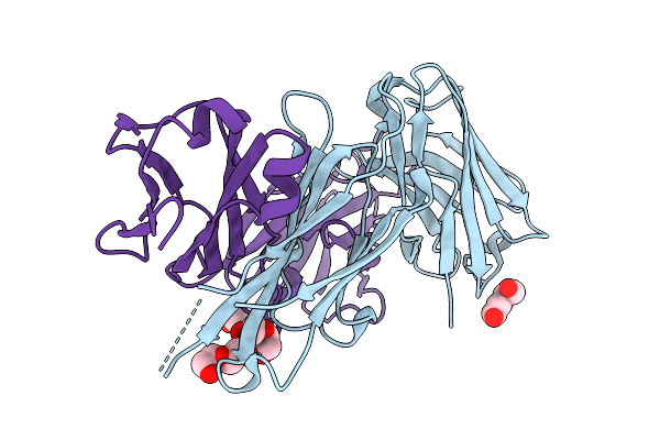 |
Organism: Homo sapiens
Method: X-RAY DIFFRACTION Release Date: 2025-10-22 Classification: IMMUNE SYSTEM Ligands: PEG |
 |
Cryo- Em Structure Of Large Subunit (Lsu) Of 75S Ribosome With P- Trna From Entamoeba Histolytica
Organism: Entamoeba histolytica hm-1:imss
Method: ELECTRON MICROSCOPY Release Date: 2025-10-15 Classification: RIBOSOME |
 |
Cryo- Em Structure Of 75S Ribosome With A/P- & P/E- Trnas From Entamoeba Histolytica
Organism: Entamoeba histolytica hm-1:imss
Method: ELECTRON MICROSCOPY Release Date: 2025-10-15 Classification: RIBOSOME |
 |
Cryo- Em Structure Of Large Subunit (Lsu) Of 75S Ribosome With A/P- & P/E- Trnas From Entamoeba Histolytica
Organism: Entamoeba histolytica hm-1:imss
Method: ELECTRON MICROSCOPY Release Date: 2025-10-15 Classification: RIBOSOME |
 |
Cryo- Em Structure Of Ribosomal Large Subunit (Lsu) From Entamoeba Histolytica At 2.8 Angstrom Resolution
Organism: Entamoeba histolytica hm-1:imss
Method: ELECTRON MICROSCOPY Release Date: 2025-10-08 Classification: RIBOSOME |
 |
Cryo- Em Structure Of Small Subunit (Body) Of 75S Ribosome With P- Trna From Entamoeba Histolytica
Organism: Entamoeba histolytica hm-1:imss
Method: ELECTRON MICROSCOPY Release Date: 2025-10-08 Classification: RIBOSOME |
 |
Cryo- Em Structure Of Small Subunit (Head) Of 75S Ribosome With P- Trna From Entamoeba Histolytica
Organism: Entamoeba histolytica hm-1:imss
Method: ELECTRON MICROSCOPY Release Date: 2025-10-08 Classification: RIBOSOME |
 |
Organism: Entamoeba histolytica hm-1:imss
Method: ELECTRON MICROSCOPY Release Date: 2025-10-08 Classification: RIBOSOME |
 |
Cryo- Em Structure Of 75S Ribosome With A/P- & P/E- Trnas From Entamoeba Histolytica Bound To Antibiotic Paromomycin
Organism: Entamoeba histolytica hm-1:imss
Method: ELECTRON MICROSCOPY Release Date: 2025-10-08 Classification: RIBOSOME Ligands: PAR |
 |
Cryo- Em Structure Of Small Subunit (Body) Of 75S Ribosome With A/P- & P/E- Trnas From Entamoeba Histolytica
Organism: Entamoeba histolytica hm-1:imss
Method: ELECTRON MICROSCOPY Release Date: 2025-10-08 Classification: RIBOSOME |
 |
Cryo- Em Structure Of Small Subunit (Head) Of 75S Ribosome With A/P- & P/E- Trnas From Entamoeba Histolytica
Organism: Entamoeba histolytica hm-1:imss
Method: ELECTRON MICROSCOPY Release Date: 2025-10-08 Classification: RIBOSOME |
 |
Cryo- Em Structure Of Small Subunit (Body) Of 75S Ribosome With A/P- & P/E- Trnas From Entamoeba Histolytica Bound To Antibiotic Paromomycin
Organism: Entamoeba histolytica hm-1:imss
Method: ELECTRON MICROSCOPY Release Date: 2025-10-08 Classification: RIBOSOME Ligands: PAR |
 |
Cryoem Structure Of Respiratory Syncytial Virus Polymerase In Complex With Novel Non-Nucleoside Inhibitor Compound 16
Organism: Human respiratory syncytial virus, Synthetic construct
Method: ELECTRON MICROSCOPY Release Date: 2025-09-24 Classification: VIRAL PROTEIN,TRANSFERASE/INHIBITOR |
 |
Cryoem Map Of Respiratory Syncytial Virus Polymerase With Non-Nucleoside Inhibitor Compound 21
Organism: Human respiratory syncytial virus, Synthetic construct
Method: ELECTRON MICROSCOPY Release Date: 2025-09-24 Classification: VIRAL PROTEIN/INHIBITOR |
 |
Organism: Severe acute respiratory syndrome coronavirus 2
Method: X-RAY DIFFRACTION Release Date: 2025-09-24 Classification: VIRAL PROTEIN Ligands: A1MA2, EDO |
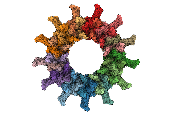 |
In Situ Cryo-Em Structure Of Outer Membrane Cap (Omc) Of The Legionella Dot/Icm T4Ss Machine
Organism: Legionella pneumophila subsp. pneumophila
Method: ELECTRON MICROSCOPY Release Date: 2025-09-17 Classification: PROTEIN TRANSPORT |