
Search Count: 23
 |
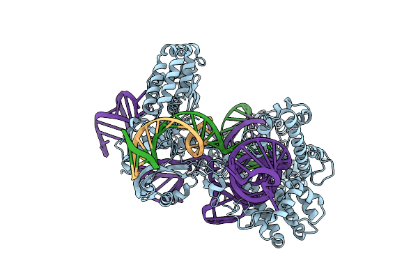
Organism: Streptococcus pyogenes, Synthetic construct
Method: ELECTRON MICROSCOPY Release Date: 2025-10-22 Classification: IMMUNE SYSTEM/DNA/RNA |
 |
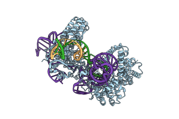
Organism: Streptococcus pyogenes, Synthetic construct
Method: ELECTRON MICROSCOPY Release Date: 2025-10-22 Classification: IMMUNE SYSTEM/DNA/RNA |
 |
Organism: Streptococcus pyogenes, Synthetic construct
Method: ELECTRON MICROSCOPY Release Date: 2025-10-22 Classification: IMMUNE SYSTEM/DNA/RNA |
 |
Organism: Streptococcus pyogenes, Synthetic construct
Method: ELECTRON MICROSCOPY Release Date: 2025-10-22 Classification: IMMUNE SYSTEM/DNA/RNA |
 |
Organism: Streptococcus pyogenes, Synthetic construct
Method: ELECTRON MICROSCOPY Release Date: 2025-10-22 Classification: IMMUNE SYSTEM/DNA/RNA |
 |
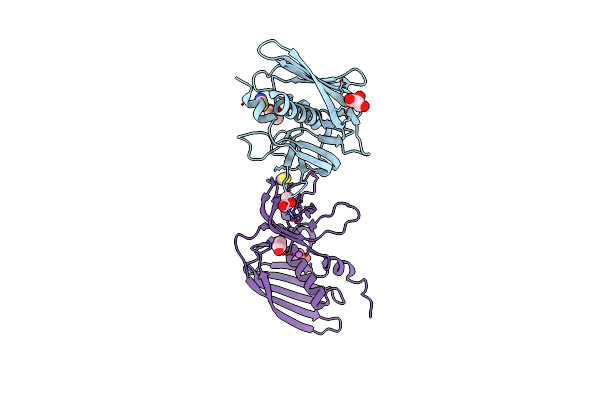
Crystal Structure Of The Glycoprotein Vi Loop Truncation Mutant Pavs-Papykn
Organism: Homo sapiens
Method: X-RAY DIFFRACTION Resolution:1.90 Å Release Date: 2018-09-05 Classification: BLOOD CLOTTING Ligands: CL, PO4, PG6, NAG |
 |
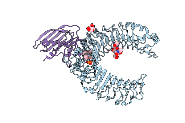
Crystal Structure Of Glycoprotein Vi In Complex With Collagen-Peptide (Gpo)5
Organism: Homo sapiens
Method: X-RAY DIFFRACTION Resolution:2.50 Å Release Date: 2018-09-05 Classification: BLOOD CLOTTING Ligands: CL, NAG, PG4 |
 |
Crystal Structure Of Glycoprotein Vi In Complex With Collagen-Peptide (Gpo)3
Organism: Homo sapiens
Method: X-RAY DIFFRACTION Resolution:2.50 Å Release Date: 2018-09-05 Classification: BLOOD CLOTTING Ligands: NAG, PG4, CL |
 |
Crystal Structure Of Human Beta1-Coronavirus Oc43 Nl/A/2005 Hemagglutinin-Esterase
Organism: Human coronavirus oc43
Method: X-RAY DIFFRACTION Resolution:2.45 Å Release Date: 2017-03-22 Classification: VIRAL PROTEIN Ligands: NAG, ACY |
 |
Crystal Structure Of Rat Coronavirus Strain New-Jersey Hemagglutinin-Esterase
Organism: Rat coronavirus (strain nj)
Method: X-RAY DIFFRACTION Resolution:2.20 Å Release Date: 2016-05-11 Classification: VIRAL PROTEIN Ligands: NAG, NA, ZN |
 |
Crystal Structure Of Mouse Hepatitis Virus Strain Dvim Hemagglutinin-Esterase
Organism: Murine coronavirus
Method: X-RAY DIFFRACTION Resolution:2.00 Å Release Date: 2016-05-11 Classification: VIRAL PROTEIN |
 |
Crystal Structure Of Rat Coronavirus Strain New-Jersey Hemagglutinin-Esterase In Complex With 4N-Acetyl Sialic Acid
Organism: Rat coronavirus
Method: X-RAY DIFFRACTION Resolution:1.85 Å Release Date: 2016-05-11 Classification: VIRAL PROTEIN Ligands: NAG, NA, ZN, 6KL |
 |
Organism: Staphylococcus aureus (strain nctc 8325)
Method: X-RAY DIFFRACTION Resolution:1.94 Å Release Date: 2015-08-19 Classification: IMMUNE SYSTEM Ligands: CL, SCN, GOL, NA |
 |
Organism: Mus musculus, Staphylococcus aureus (strain nctc 8325)
Method: X-RAY DIFFRACTION Resolution:3.20 Å Release Date: 2015-08-19 Classification: IMMUNE SYSTEM Ligands: NAG, PCW, CL |
 |
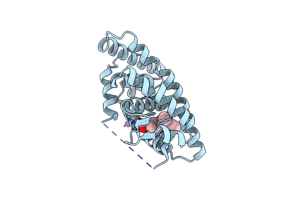
Crystal Structure Of Human Retinoid X Receptor Alpha-Ligand Binding Domain Complex With 8-Methyl Uab30 And The Coactivator Peptide Grip-1
Organism: Homo sapiens
Method: X-RAY DIFFRACTION Resolution:2.30 Å Release Date: 2014-06-18 Classification: TRANSCRIPTION Ligands: 2VR |
 |
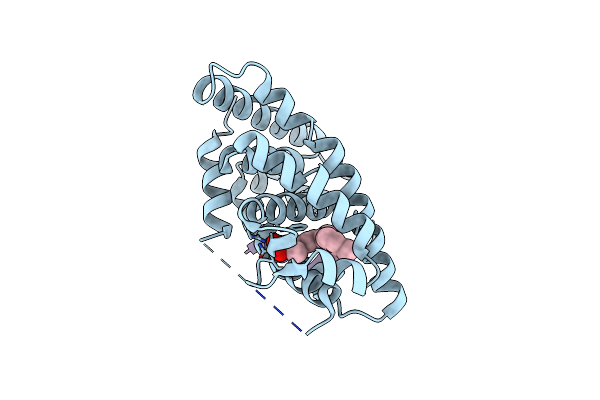
Crystal Structure Of Human Retinoid X Receptor Alpha-Ligand Binding Domain Complex With 7-Methyl Uab30 And The Coactivator Peptide Grip-1
Organism: Homo sapiens
Method: X-RAY DIFFRACTION Resolution:2.00 Å Release Date: 2014-06-18 Classification: TRANSCRIPTION Ligands: 2VP |
 |
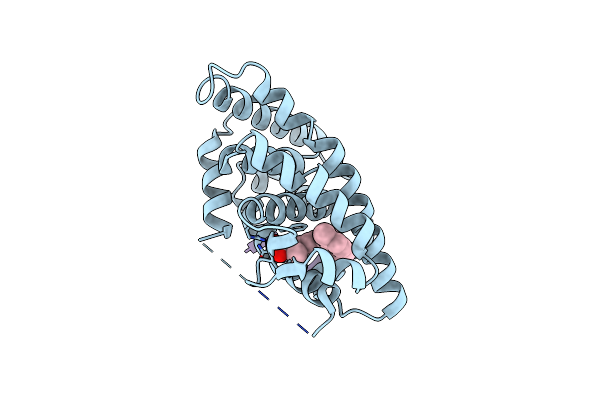
Crystal Structure Of Human Retinoid X Receptor Alpha-Ligand Binding Domain Complex With 6-Methyl Uab30 And The Coactivator Peptide Grip-1
Organism: Homo sapiens
Method: X-RAY DIFFRACTION Resolution:2.00 Å Release Date: 2014-06-18 Classification: TRANSCRIPTION Ligands: 2VZ |
 |
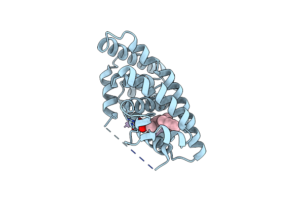
Crystal Structure Of Human Retinoid X Receptor Alpha-Ligand Binding Domain Complex With 5-Methyl Uab30 And The Coactivator Peptide Grip-1
Organism: Homo sapiens
Method: X-RAY DIFFRACTION Resolution:2.00 Å Release Date: 2014-06-18 Classification: TRANSCRIPTION Ligands: 2W0 |
 |
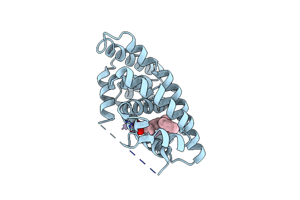
Crystal Structure Of Human Retinoid X Receptor Alpha-Ligand Binding Domain Complex With (S) 4-Methyl 9Cuab30 Coactivator Peptide Grip-1
Organism: Homo sapiens
Method: X-RAY DIFFRACTION Resolution:2.40 Å Release Date: 2014-01-22 Classification: TRANSCRIPTION Ligands: 29V |
 |
Crystal Structure Of Human Retinoid X Receptor Alpha-Ligand Binding Domain Complex With (R)4-Methyl 9Cuab30 And Coactivator Peptide Grip-1
Organism: Homo sapiens
Method: X-RAY DIFFRACTION Resolution:2.20 Å Release Date: 2014-01-22 Classification: TRANSCRIPTION Ligands: R4M |