
Search Count: 12
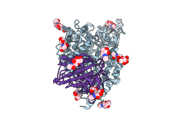 |
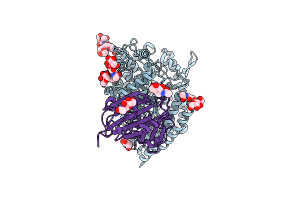
Structure Of The Rattus Norvegicus Ace2 Receptor Bound Hsitaly2011 Rbd Complex
Organism: Rattus norvegicus, Merbecovirus
Method: ELECTRON MICROSCOPY Release Date: 2025-08-27 Classification: HYDROLASE/VIRAL PROTEIN Ligands: NAG, ZN |
 |
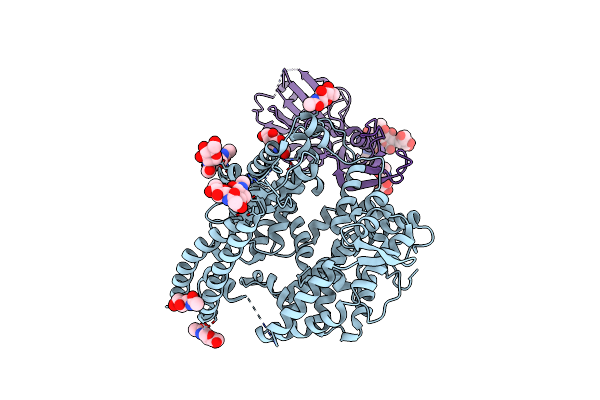
Organism: Eptesicus fuscus, Merbecovirus
Method: ELECTRON MICROSCOPY Release Date: 2025-08-27 Classification: HYDROLASE/VIRAL PROTEIN Ligands: NAG, ZN |
 |
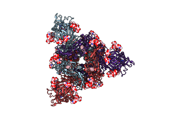
Organism: Pipistrellus bat coronavirus hku5, Homo sapiens
Method: ELECTRON MICROSCOPY Release Date: 2025-03-12 Classification: VIRAL PROTEIN/HYDROLASE Ligands: NAG |
 |
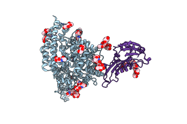
Organism: Merbecovirus, Pteronotus davyi
Method: ELECTRON MICROSCOPY Release Date: 2025-03-05 Classification: VIRAL PROTEIN Ligands: NAG |
 |
Organism: Pipistrellus bat coronavirus hku5
Method: ELECTRON MICROSCOPY Resolution:2.00 Å Release Date: 2025-02-26 Classification: VIRAL PROTEIN Ligands: NAG, ZN, FOL, EIC |
 |
Organism: Pipistrellus bat coronavirus hku5
Method: ELECTRON MICROSCOPY Resolution:2.00 Å Release Date: 2025-02-26 Classification: VIRAL PROTEIN/IMMUNE SYSTEM Ligands: NAG, FOL, EIC |
 |
Organism: Pipistrellus abramus, Pipistrellus bat coronavirus hku5
Method: ELECTRON MICROSCOPY Resolution:3.10 Å Release Date: 2025-02-19 Classification: VIRAL PROTEIN/HYDROLASE Ligands: NAG, ZN |
 |
Merbecovirus Pnnl2018B Spike Glycoprotein Rbd Bound To The P. Nathusii Ace2
Organism: Pipistrellus nathusii, Merbecovirus
Method: ELECTRON MICROSCOPY Release Date: 2025-02-19 Classification: VIRAL PROTEIN Ligands: NAG, ZN |
 |
Organism: Bos taurus, Pipistrellus bat coronavirus hku5
Method: ELECTRON MICROSCOPY Release Date: 2025-02-19 Classification: VIRAL PROTEIN/HYDROLASE Ligands: NAG, ZN |
 |
Organism: Pipistrellus nathusii, Middle east respiratory syndrome-related coronavirus
Method: ELECTRON MICROSCOPY Release Date: 2025-02-12 Classification: VIRAL PROTEIN/HYDROLASE Ligands: NAG, ZN |
 |
Organism: Severe acute respiratory syndrome coronavirus 2, Homo sapiens
Method: ELECTRON MICROSCOPY Release Date: 2024-11-20 Classification: VIRAL PROTEIN Ligands: NAG |
 |
Sars-Cov-2 S + S2L20 (Local Refinement Of Ntd And S2L20 Fab Variable Region)
Organism: Homo sapiens, Severe acute respiratory syndrome coronavirus 2
Method: ELECTRON MICROSCOPY Release Date: 2024-11-20 Classification: VIRAL PROTEIN/IMMUNE SYSTEM Ligands: NAG |