
Search Count: 10
 |
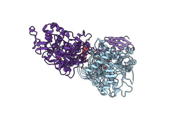
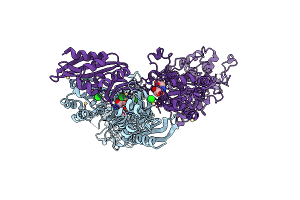
Crystal Structure Of Pbp2A From Mrsa In Complex With Piperacillin And Quinazolinone
Organism: Staphylococcus aureus (strain mu50 / atcc 700699)
Method: X-RAY DIFFRACTION Resolution:2.50 Å Release Date: 2019-11-27 Classification: HYDROLASE Ligands: QLN, JPP, CL, CD, MUR |
 |
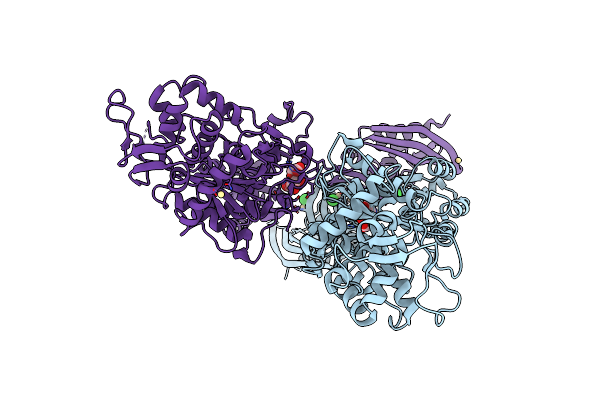
Organism: Staphylococcus aureus
Method: X-RAY DIFFRACTION Resolution:1.98 Å Release Date: 2017-02-08 Classification: Penicillin binding protein Ligands: CD, MUR |
 |
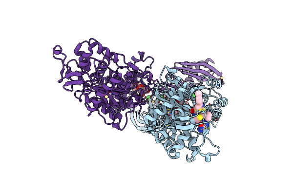
Organism: Staphylococcus aureus
Method: X-RAY DIFFRACTION Resolution:2.00 Å Release Date: 2017-02-08 Classification: Penicillin binding protein Ligands: CD, MUR |
 |
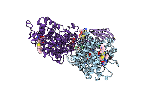
Organism: Staphylococcus aureus
Method: X-RAY DIFFRACTION Resolution:2.00 Å Release Date: 2017-02-08 Classification: Penicillin binding protein Ligands: CD, MUR |
 |
Organism: Staphylococcus aureus subsp. aureus mu50
Method: X-RAY DIFFRACTION Resolution:1.95 Å Release Date: 2015-02-11 Classification: HYDROLASE Ligands: CD, CL, MUR, QNZ |
 |
Organism: Staphylococcus aureus (strain mu50 / atcc 700699)
Method: X-RAY DIFFRACTION Resolution:2.35 Å Release Date: 2014-09-10 Classification: HYDROLASE Ligands: CD, CL, MUR |
 |
Organism: Staphylococcus aureus
Method: X-RAY DIFFRACTION Resolution:3.00 Å Release Date: 2014-05-21 Classification: HYDROLASE Ligands: CD, CL, MUR |
 |
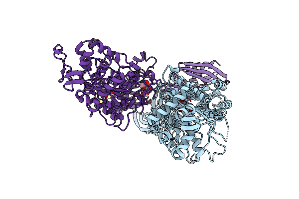
Crystal Structure Of Ceftaroline Acyl-Pbp2A From Mrsa With Non- Covalently Bound Ceftaroline And Muramic Acid At Allosteric Site Obtained By Soaking
Organism: Staphylococcus aureus subsp. aureus mu50
Method: X-RAY DIFFRACTION Resolution:2.25 Å Release Date: 2013-10-09 Classification: HYDROLASE Ligands: CD, CL, AI8, 1W8, MUR |
 |
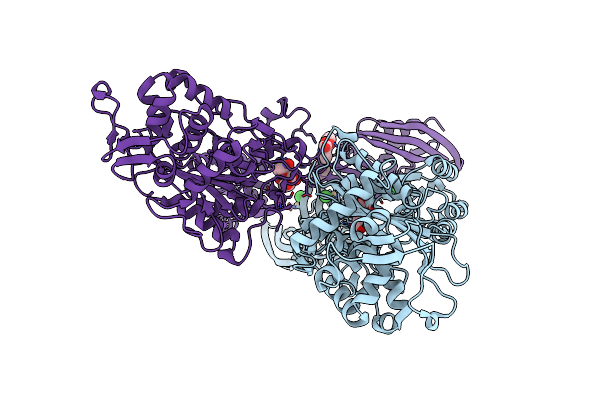
Crystal Structure Of Ceftaroline Acyl-Pbp2A From Mrsa With Non- Covalently Bound Ceftaroline And Muramic Acid At Allosteric Site Obtained By Cocrystallization
Organism: Staphylococcus aureus subsp. aureus mu50
Method: X-RAY DIFFRACTION Resolution:2.60 Å Release Date: 2013-10-09 Classification: HYDROLASE Ligands: AI8, CD, CL, 1W8, MUR |
 |
Interaction Of A Legume Lectin With Two Components Of The Bacterial Cell Wall
Organism: Lathyrus ochrus
Method: X-RAY DIFFRACTION Resolution:2.05 Å Release Date: 1994-04-30 Classification: LECTIN Ligands: CA, MN, MUR |