
Search Count: 64
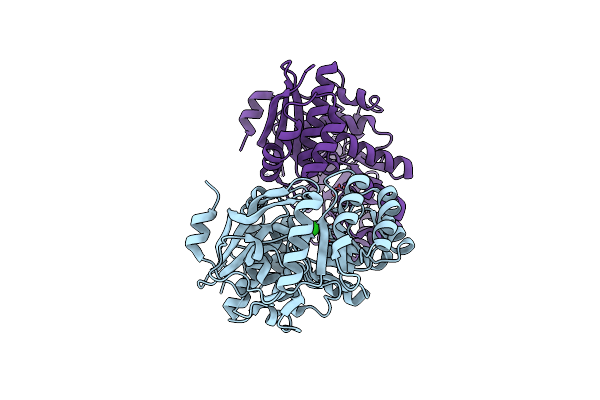 |
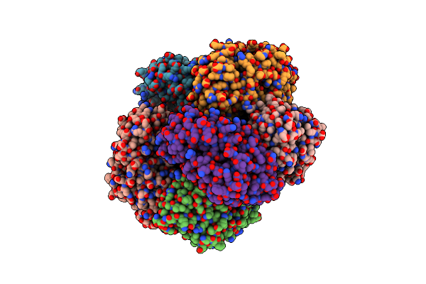
Crystal Structure Of 2-Chloromuconate Cycloisomerase From Rhodococcus Opacus 1Cp
Organism: Rhodococcus opacus
Method: X-RAY DIFFRACTION Resolution:2.70 Å Release Date: 2014-07-09 Classification: ISOMERASE Ligands: MN, CL |
 |
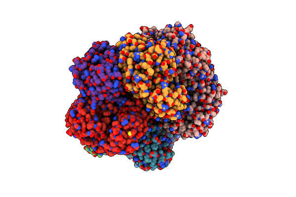
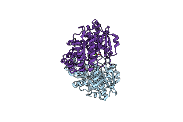
Crystal Structure Of Dipeptide Epimerase From Methylococcus Capsulatus Complexed With Mg Ion
Organism: Methylococcus capsulatus
Method: X-RAY DIFFRACTION Resolution:2.20 Å Release Date: 2011-05-11 Classification: ISOMERASE Ligands: MG, SO4, GOL |
 |
Crystal Structure Of Dipeptide Epimerase From Methylococcus Capsulatus Complexed With Mg And Dipeptide L-Arg-D-Lys
Organism: Methylococcus capsulatus
Method: X-RAY DIFFRACTION Resolution:2.70 Å Release Date: 2011-04-27 Classification: ISOMERASE Ligands: SO4, ARG, DLY, MG, 1PE |
 |
Crystal Structure Of Dipeptide Epimerase From Cytophaga Hutchinsonii Complexed With Mg And Dipeptide D-Ala-L-Val
Organism: Cytophaga hutchinsonii
Method: X-RAY DIFFRACTION Resolution:3.00 Å Release Date: 2011-02-16 Classification: ISOMERASE Ligands: MG, DAL, VAL |
 |
Crystal Structure Of Dipeptide Epimerase From Cytophaga Hutchinsonii Complexed With Mg And Dipeptide D-Ala-L-Ala
Organism: Cytophaga hutchinsonii
Method: X-RAY DIFFRACTION Resolution:3.00 Å Release Date: 2011-02-16 Classification: ISOMERASE Ligands: MG, DAL, ALA |
 |
Structure Of Dipeptide Epimerase From Bacteroides Thetaiotaomicron Complexed With L-Ala-D-Glu; Nonproductive Substrate Binding.
Organism: Bacteroides thetaiotaomicron
Method: X-RAY DIFFRACTION Resolution:1.60 Å Release Date: 2010-07-21 Classification: ISOMERASE Ligands: ALA, DGL, MG |
 |
Structure Of Dipeptide Epimerase From Bacteroides Thetaiotaomicron Complexed With L-Pro-D-Glu; Nonproductive Substrate Binding.
Organism: Bacteroides thetaiotaomicron
Method: X-RAY DIFFRACTION Resolution:1.50 Å Release Date: 2010-07-21 Classification: ISOMERASE Ligands: PRO, DGL, MG |
 |
Structure Of Dipeptide Epimerase From Bacteroides Thetaiotaomicron Complexed With L-Ala-D-Glu; Productive Substrate Binding.
Organism: Bacteroides thetaiotaomicron
Method: X-RAY DIFFRACTION Resolution:2.00 Å Release Date: 2010-07-21 Classification: ISOMERASE Ligands: ALA, DGL, MG, SO4 |
 |
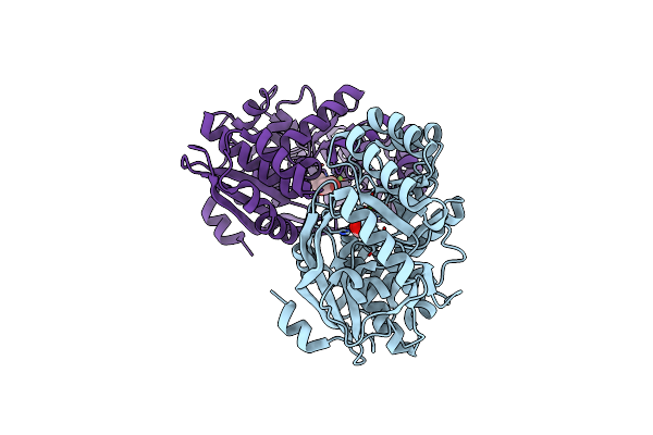
Crystal Structure Of A Muconate Cycloisomerase From Azorhizobium Caulinodans
Organism: Azorhizobium caulinodans
Method: X-RAY DIFFRACTION Resolution:2.20 Å Release Date: 2010-07-07 Classification: ISOMERASE Ligands: MG, GOL |
 |
Crystal Structure Of The Mutant Y90F Of Divergent Galactarate Dehydratase From Oceanobacillus Iheyensis Complexed With Mg And Galactarate
Organism: Oceanobacillus iheyensis hte831
Method: X-RAY DIFFRACTION Resolution:1.80 Å Release Date: 2009-12-22 Classification: ISOMERASE Ligands: GAE, MG |
 |
Organism: Jannaschia sp.
Method: X-RAY DIFFRACTION Resolution:1.90 Å Release Date: 2009-07-21 Classification: ISOMERASE Ligands: K, MG |
 |
Crystal Structure Of Muconate Lactonizing Enzyme From Corynebacterium Glutamicum
Organism: Corynebacterium glutamicum
Method: X-RAY DIFFRACTION Resolution:2.20 Å Release Date: 2009-07-14 Classification: ISOMERASE Ligands: ACY, MG |
 |
Organism: Ruegeria pomeroyi
Method: X-RAY DIFFRACTION Resolution:1.70 Å Release Date: 2009-07-14 Classification: ISOMERASE Ligands: MG, NA |
 |
Crystal Structure Of Muconate Lactonizing Enzyme From Pseudomonas Fluorescens Complexed With Muconolactone
Organism: Pseudomonas fluorescens
Method: X-RAY DIFFRACTION Resolution:1.80 Å Release Date: 2009-03-31 Classification: ISOMERASE Ligands: MUC, MG |
 |
Crystal Structure Of Muconate Lactonizing Enzyme From Mucobacterium Smegmatis
Organism: Mycobacterium smegmatis
Method: X-RAY DIFFRACTION Resolution:1.60 Å Release Date: 2009-03-03 Classification: ISOMERASE Ligands: MG |
 |
Crystal Structure Of Muconate Lactonizing Enzyme From Mucobacterium Smegmatis Complexed With Muconolactone
Organism: Mycobacterium smegmatis
Method: X-RAY DIFFRACTION Resolution:1.60 Å Release Date: 2009-03-03 Classification: ISOMERASE Ligands: MG, MUC |
 |
Crystal Structure Of Muconate Lactonizing Enzyme From Mucobacterium Smegmatis Complexed With Muconolactone
Organism: Mycobacterium smegmatis
Method: X-RAY DIFFRACTION Resolution:2.00 Å Release Date: 2009-03-03 Classification: ISOMERASE Ligands: MG, MUC |
 |
Crystal Structure Of Muconate Lactonizing Enzyme From Pseudomonas Fluorescens Complexed With Muconolactone
Organism: Pseudomonas fluorescens
Method: X-RAY DIFFRACTION Resolution:1.70 Å Release Date: 2009-03-03 Classification: ISOMERASE Ligands: MG, MUC |
 |
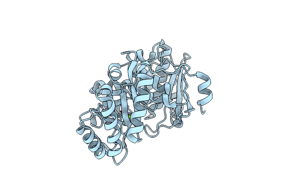
Crystal Structure Of Divergent Enolase From Oceanobacillus Iheyensis Complexed With Phosphate
Organism: Oceanobacillus iheyensis hte831
Method: X-RAY DIFFRACTION Resolution:1.60 Å Release Date: 2009-03-03 Classification: ISOMERASE, LYASE Ligands: PO4 |
 |
Crystal Structure Of Divergent Enolase From Oceanobacillus Iheyensis Complexed With Mg
Organism: Oceanobacillus iheyensis hte831
Method: X-RAY DIFFRACTION Resolution:1.80 Å Release Date: 2009-02-03 Classification: ISOMERASE Ligands: MG |