
Search Count: 476
All
Selected
 |
Organism: Litorilinea aerophila
Method: ELECTRON MICROSCOPY Release Date: 2026-01-28 Classification: PROTEIN FIBRIL |
 |
Organism: Litorilinea aerophila
Method: ELECTRON MICROSCOPY Release Date: 2026-01-28 Classification: PROTEIN FIBRIL |
 |
Organism: Homo sapiens
Method: ELECTRON MICROSCOPY Release Date: 2026-01-21 Classification: STRUCTURAL PROTEIN |
 |
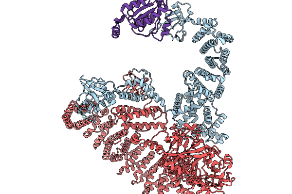
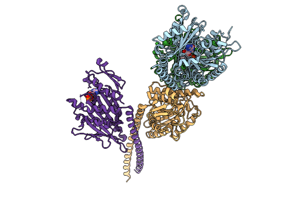
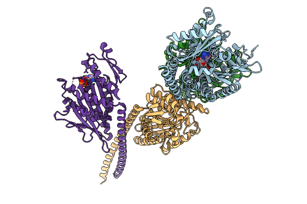
Cryo-Em Structure Of The Cytosolic Armh2-Efcab9-Catsperz Subcomplex Of The Mouse Catspermasome
Organism: Mus musculus
Method: ELECTRON MICROSCOPY Release Date: 2025-12-31 Classification: CYTOSOLIC PROTEIN |
 |
Organism: Pseudomonas aeruginosa
Method: X-RAY DIFFRACTION Resolution:4.54 Å Release Date: 2025-12-10 Classification: MOTOR PROTEIN |
 |
Organism: Homo sapiens, Bos taurus
Method: ELECTRON MICROSCOPY Release Date: 2025-11-19 Classification: PROTEIN BINDING Ligands: ATP, MG |
 |
Organism: Homo sapiens
Method: ELECTRON MICROSCOPY Resolution:6.98 Å Release Date: 2025-11-19 Classification: SIGNALING PROTEIN |
 |
Organism: Homo sapiens
Method: ELECTRON MICROSCOPY Resolution:7.52 Å Release Date: 2025-11-19 Classification: SIGNALING PROTEIN |
 |
Organism: Drosophila melanogaster, Sus scrofa
Method: ELECTRON MICROSCOPY Release Date: 2025-10-15 Classification: CELL CYCLE Ligands: GTP, GDP, ADP, MG, ALF |
 |
Organism: Campylobacter jejuni subsp. jejuni serotype o:23/36 (strain 81-176)
Method: ELECTRON MICROSCOPY Release Date: 2025-10-15 Classification: STRUCTURAL PROTEIN |
 |
Organism: Drosophila melanogaster, Sus scrofa
Method: ELECTRON MICROSCOPY Release Date: 2025-10-08 Classification: CELL CYCLE Ligands: GTP, GDP, MG, ADP |
 |
Organism: Drosophila melanogaster, Sus scrofa
Method: ELECTRON MICROSCOPY Release Date: 2025-10-08 Classification: CELL CYCLE Ligands: GTP, GDP, MG, ADP |
 |
Organism: Drosophila melanogaster, Sus scrofa
Method: ELECTRON MICROSCOPY Release Date: 2025-10-08 Classification: CELL CYCLE Ligands: GTP, GDP, ADP, MG, ALF |
 |
Organism: Litorilinea aerophila
Method: ELECTRON MICROSCOPY Release Date: 2025-07-23 Classification: PROTEIN FIBRIL |
 |
Organism: Mus musculus
Method: X-RAY DIFFRACTION Resolution:2.45 Å Release Date: 2025-07-16 Classification: CYTOSOLIC PROTEIN |
 |
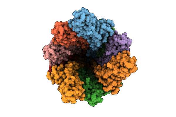
Structure Of P. Gingivalis Pork And Porn Complexes From Cryo Electron Microscopy
Organism: Porphyromonas gingivalis w83
Method: ELECTRON MICROSCOPY Resolution:3.57 Å Release Date: 2025-07-16 Classification: TRANSPORT PROTEIN Ligands: MAN, CA |
 |
Structure Of P. Gingivalis Pork And Porn Complexes From Cryo Electron Microscopy
Organism: Porphyromonas gingivalis w83
Method: ELECTRON MICROSCOPY Resolution:2.75 Å Release Date: 2025-07-16 Classification: TRANSPORT PROTEIN Ligands: BMA, CA |
 |
Organism: Myxococcus xanthus dk 1622
Method: X-RAY DIFFRACTION Resolution:1.89 Å Release Date: 2025-06-04 Classification: SIGNALING PROTEIN Ligands: EDO |
 |
Organism: Litorilinea aerophila
Method: ELECTRON MICROSCOPY Release Date: 2025-05-21 Classification: PROTEIN FIBRIL |
 |
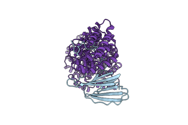
Crystal Structure Of Actin Capping Protein In Complex With A Fragment Of Legionella Pneumophila Ravb
Organism: Gallus gallus, Legionella pneumophila subsp. pneumophila (strain philadelphia 1 / atcc 33152 / dsm 7513)
Method: X-RAY DIFFRACTION Resolution:2.00 Å Release Date: 2025-05-07 Classification: PROTEIN BINDING Ligands: FMT |