
Search Count: 27
 |
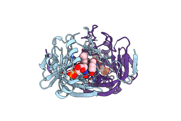
Organism: Clostridium cochlearium
Method: X-RAY DIFFRACTION Resolution:1.82 Å Release Date: 2019-08-14 Classification: ISOMERASE Ligands: B12, FWK, TAR, GOL |
 |
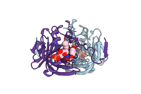
Organism: Clostridium cochlearium
Method: X-RAY DIFFRACTION Resolution:2.10 Å Release Date: 2019-08-14 Classification: ISOMERASE Ligands: B12, 8ZB, TAR |
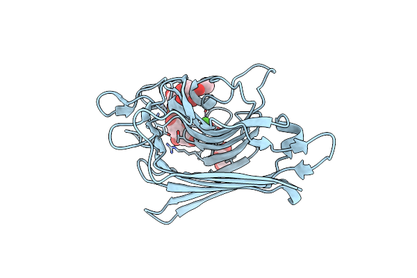 |
Crystal Structure Of A Recombinant Vatairea Macrocarpa Seed Lectin Complexed With Galnac
Organism: Vatairea macrocarpa
Method: X-RAY DIFFRACTION Resolution:2.70 Å Release Date: 2016-01-27 Classification: SUGAR BINDING PROTEIN Ligands: CA, MN, A2G, CIT, GOL, SO4 |
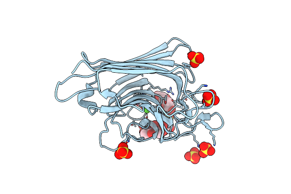 |
Crystal Structure Of A Recombinant Vatairea Macrocarpa Seed Lectin Complexed With Tn Antigen
Organism: Vatairea macrocarpa
Method: X-RAY DIFFRACTION Resolution:1.97 Å Release Date: 2016-01-27 Classification: SUGAR BINDING PROTEIN Ligands: CA, MN, CIT, SO4, SER, A2G |
 |
Organism: Vatairea macrocarpa
Method: X-RAY DIFFRACTION Resolution:1.90 Å Release Date: 2016-01-27 Classification: SUGAR BINDING PROTEIN Ligands: CA, MN, GOL, 1PE |
 |
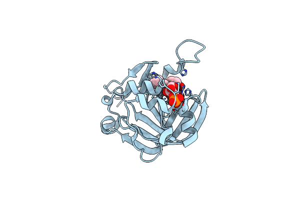
Organism: Clostridium botulinum
Method: X-RAY DIFFRACTION Resolution:1.20 Å Release Date: 2015-11-11 Classification: HYDROLASE Ligands: ZN, K |
 |
Organism: Enterobacter sp.
Method: X-RAY DIFFRACTION Resolution:1.49 Å Release Date: 2015-06-10 Classification: LYASE Ligands: PO4, GLY, SO4 |
 |
Organism: Enterobacter sp.
Method: X-RAY DIFFRACTION Resolution:1.15 Å Release Date: 2015-06-10 Classification: LYASE |
 |
Crystal Structure Of Fmn-Binding Protein (Np_142786.1) From Pyrococcus Horikoshii With Bound P-Hydroxybenzaldehyde
Organism: Pyrococcus horikoshii
Method: X-RAY DIFFRACTION Resolution:2.10 Å Release Date: 2014-05-14 Classification: FMN-BINDING PROTEIN Ligands: FMN, HBA |
 |
Crystal Structure Of Fmn-Binding Protein (Np_142786.1) From Pyrococcus Horikoshii With Bound Benzene-1,4-Diol
Organism: Pyrococcus horikoshii
Method: X-RAY DIFFRACTION Resolution:1.68 Å Release Date: 2014-05-14 Classification: FMN-BINDING PROTEIN Ligands: FMN, HQE |
 |
Crystal Structure Of Fmn-Binding Protein (Yp_005476) From Thermus Thermophilus With Bound P-Hydroxybenzaldehyde
Organism: Thermus thermophilus
Method: X-RAY DIFFRACTION Resolution:1.85 Å Release Date: 2014-05-14 Classification: FMN-BINDING PROTEIN Ligands: HBA, FMN |
 |
Crystal Structure Of Fmn-Binding Protein (Yp_005476) From Thermus Thermophilus With Bound Benzene-1,4-Diol
Organism: Thermus thermophilus
Method: X-RAY DIFFRACTION Resolution:2.15 Å Release Date: 2014-05-14 Classification: FMN-BINDING PROTEIN Ligands: HQE, FMN |
 |
Crystal Structure Of Fmn-Binding Protein (Np_142786.1) From Pyrococcus Horikoshii With Bound 1-Cyclohex-2-Enone
Organism: Pyrococcus horikoshii
Method: X-RAY DIFFRACTION Resolution:1.75 Å Release Date: 2014-05-14 Classification: OXIDOREDUCTASE Ligands: FMN, A2Q |
 |
Crystal Structure Of Fmn-Binding Protein (Yp_005476) From Thermus Thermophilus With Bound 1-Cyclohex-2-Enone
Organism: Thermus thermophilus
Method: X-RAY DIFFRACTION Resolution:1.65 Å Release Date: 2014-05-14 Classification: FMN-BINDING PROTEIN Ligands: A2Q, FMN |
 |
First Crystal Structure Of An (R)-Selective Omega-Transaminase From Aspergillus Terreus
Organism: Aspergillus terreus
Method: X-RAY DIFFRACTION Resolution:1.63 Å Release Date: 2014-02-12 Classification: TRANSFERASE Ligands: PLP, PDG, CL, CA |
 |
Biochemical And Structural Characterisation Of A Novel Manganese- Dependent Hydroxynitrile Lyase From Bacteria
Organism: Granulicella tundricola
Method: X-RAY DIFFRACTION Resolution:2.46 Å Release Date: 2013-09-11 Classification: LYASE Ligands: MN |
 |
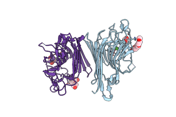
Crystal Structure Of An Immunoglobulin-Binding Fragment Of The Trimeric Autotransporter Adhesin Eibd
Organism: Escherichia coli
Method: X-RAY DIFFRACTION Resolution:1.99 Å Release Date: 2011-07-20 Classification: CELL ADHESION Ligands: CL |
 |
Escherichia Coli Immunoglobulin-Binding Protein Eibd 391-438 Fused To Gcn4 Adaptors
Organism: Enterobacteria phage p-eibd
Method: X-RAY DIFFRACTION Resolution:2.80 Å Release Date: 2011-07-20 Classification: CELL ADHESION Ligands: CL |
 |
Organism: Rhodococcus ruber
Method: X-RAY DIFFRACTION Resolution:2.00 Å Release Date: 2010-08-25 Classification: OXIDOREDUCTASE Ligands: ZN, NAD, ACY, MPD |
 |
Alcohol Dehydrogenase Adh-'A' From Rhodococcus Ruber Dsm 44541 At Ph 8.5 In Complex With Nad And Butane-1,4-Diol
Organism: Rhodococcus ruber
Method: X-RAY DIFFRACTION Resolution:2.80 Å Release Date: 2010-08-11 Classification: OXIDOREDUCTASE Ligands: NAD, ZN, BU1 |