
Search Count: 219
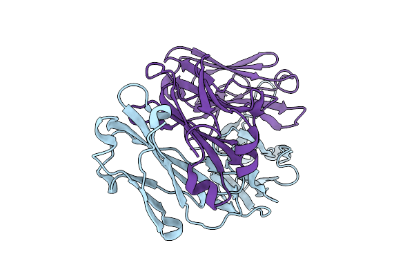 |
Organism: Homo sapiens
Method: X-RAY DIFFRACTION Release Date: 2025-12-03 Classification: IMMUNE SYSTEM |
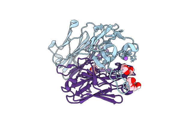 |
Organism: Homo sapiens
Method: X-RAY DIFFRACTION Release Date: 2025-12-03 Classification: IMMUNE SYSTEM Ligands: PGE, NA, PEG |
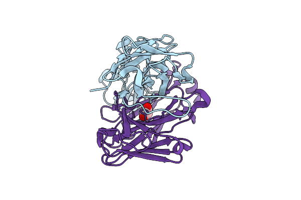 |
Organism: Homo sapiens
Method: X-RAY DIFFRACTION Release Date: 2025-12-03 Classification: IMMUNE SYSTEM Ligands: GOL |
 |
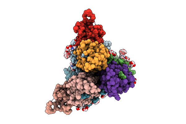
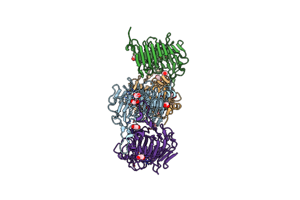
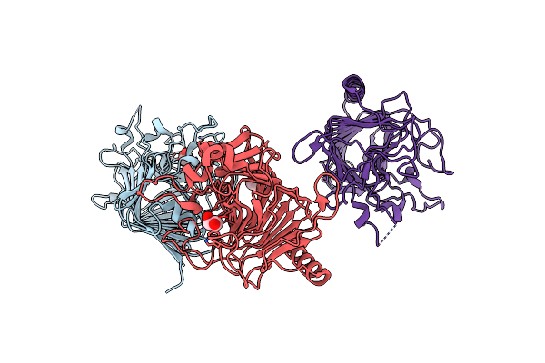
Cryoem Structure Of De Novo Antibody Fragment Scfv 6 With C. Difficile Toxin B (Tcdb)
Organism: Clostridioides difficile, Synthetic construct
Method: ELECTRON MICROSCOPY Release Date: 2025-11-12 Classification: DE NOVO PROTEIN |
 |
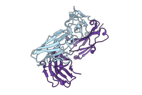
Cryoem Structure Of De Novo Vhh, Vhh_Flu_01, Bound To Influenza Ha, Strain A/Usa:Iowa/1943 H1N1.
Organism: Synthetic construct, Influenza a virus (strain a/usa:iowa/1943 h1n1)
Method: ELECTRON MICROSCOPY Release Date: 2025-11-12 Classification: DE NOVO PROTEIN Ligands: NAG |
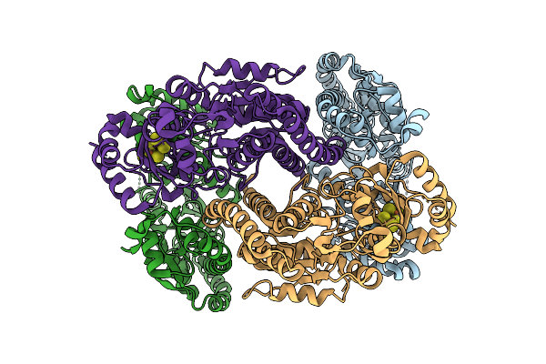 |
Rhodospirillum Rubrum Nitrogenase-Like Methylthio-Alkane Reductase Complex With An Oxidized P-Cluster
Organism: Rhodospirillum rubrum atcc 11170
Method: ELECTRON MICROSCOPY Release Date: 2025-11-05 Classification: OXIDOREDUCTASE Ligands: CLF |
 |
Organism: Mus musculus
Method: X-RAY DIFFRACTION Release Date: 2025-08-27 Classification: IMMUNE SYSTEM |
 |
Organism: Synthetic construct, Homo sapiens
Method: X-RAY DIFFRACTION Release Date: 2025-08-13 Classification: DE NOVO PROTEIN |
 |
Organism: Homo sapiens, Synthetic construct
Method: X-RAY DIFFRACTION Release Date: 2025-08-13 Classification: DE NOVO PROTEIN |
 |
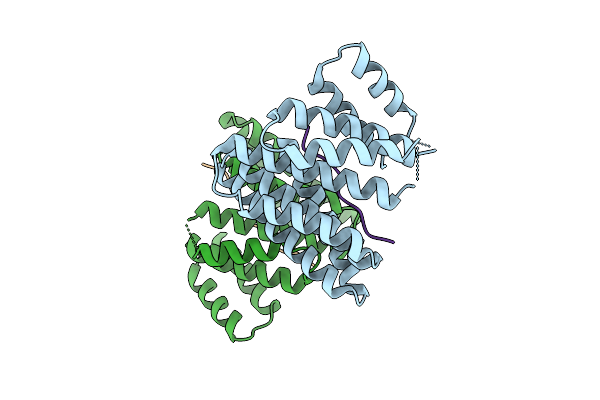
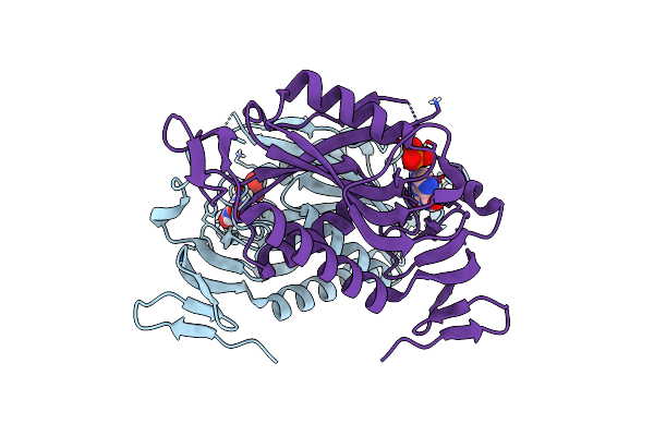
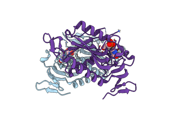
O-Glcnacase (Oga) Inhibitor Complex For The Treatment Of Alzheimer'S Disease
Organism: Homo sapiens
Method: X-RAY DIFFRACTION Resolution:2.54 Å Release Date: 2025-01-15 Classification: HYDROLASE Ligands: A1AKM |
 |
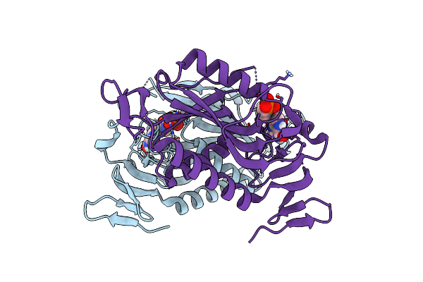
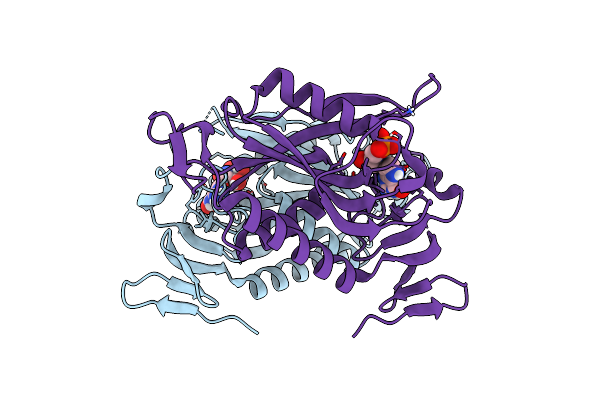
O-Glcnacase (Oga) Inhibitor Complex For The Treatment Of Alzheimer'S Disease
Organism: Homo sapiens
Method: X-RAY DIFFRACTION Resolution:2.75 Å Release Date: 2025-01-15 Classification: HYDROLASE Ligands: A1AKL |
 |
Crystal Structure Of Bacterial Pectin Methylesterase Pmec5 From B. Fibrisolvens D1T
Organism: Butyrivibrio fibrisolvens dsm 3071
Method: X-RAY DIFFRACTION Release Date: 2025-01-15 Classification: SUGAR BINDING PROTEIN Ligands: MG, NA, EDO, NH4 |
 |
Crystal Structure Of Bacterial Pectin Methylesterase Pmec2 From Rumen Butyrivibrio
Organism: Butyrivibrio fibrisolvens
Method: X-RAY DIFFRACTION Resolution:2.30 Å Release Date: 2023-08-16 Classification: SUGAR BINDING PROTEIN Ligands: CL, EDO |
 |
Crystal Structure Of Bacterial Pectin Methylesterase Pme8A From Rumen Butyrivibrio
Organism: Butyrivibrio proteoclasticus
Method: X-RAY DIFFRACTION Resolution:2.30 Å Release Date: 2023-08-16 Classification: SUGAR BINDING PROTEIN Ligands: EDO |
 |
Crystal Structure Of The Trypanosoma Cruzi Hypoxanthine-Guanine-Xanthine Phosphoribosyltransferase (Hgxprt), Isoform D, Bound To The Dead-End Complex Xanthine And Pyrophosphate
Organism: Trypanosoma cruzi (strain cl brener)
Method: X-RAY DIFFRACTION Resolution:1.54 Å Release Date: 2023-07-19 Classification: TRANSFERASE Ligands: XAN, PO4 |
 |
Crystal Structure Of The Trypanosoma Cruzi Hypoxanthine-Guanine-Xanthine Phosphoribosyltransferase (Hgxprt), Isoform D, Bound To Hydroxypropyl-Lin-Immh Phosphonate
Organism: Trypanosoma cruzi (strain cl brener)
Method: X-RAY DIFFRACTION Resolution:1.65 Å Release Date: 2023-07-19 Classification: TRANSFERASE/INHIBITOR Ligands: YCE, PO4 |
 |
Crystal Structure Of The Trypanosoma Cruzi Hypoxanthine-Guanine-Xanthine Phosphoribosyltransferase (Hgxprt), Isoform D, Bound To (S)-Serme-Immh Phosphonate
Organism: Trypanosoma cruzi (strain cl brener)
Method: X-RAY DIFFRACTION Resolution:1.52 Å Release Date: 2023-07-19 Classification: TRANSFERASE/INHIBITOR Ligands: YC9 |
 |
Crystal Structure Of The Trypanosoma Cruzi Hypoxanthine-Guanine-Xanthine Phosphoribosyltransferase (Hgxprt), Isoform D, Bound To (R)-Serme-Immh Phosphonate
Organism: Trypanosoma cruzi (strain cl brener)
Method: X-RAY DIFFRACTION Resolution:1.80 Å Release Date: 2023-07-19 Classification: TRANSFERASE/INHIBITOR Ligands: YC9 |
 |
Crystal Structure Of The Trypanosoma Cruzi Hypoxanthine-Guanine-Xanthine Phosphoribosyltransferase (Hgxprt), Isoform D, Bound To Immucillin-Hp
Organism: Trypanosoma cruzi (strain cl brener)
Method: X-RAY DIFFRACTION Resolution:1.54 Å Release Date: 2023-07-19 Classification: TRANSFERASE Ligands: IRP |
 |
Crystal Structure Of The Trypanosoma Cruzi Hypoxanthine-Guanine-Xanthine Phosphoribosyltransferase (Hgxprt), Isoform D, Bound To Immucillin-Gp, Showing The Structure Of The Complete Active Site In Its Open Conformation
Organism: Trypanosoma cruzi (strain cl brener)
Method: X-RAY DIFFRACTION Resolution:1.31 Å Release Date: 2023-07-19 Classification: TRANSFERASE/INHIBITOR Ligands: IMU |