
Search Count: 40
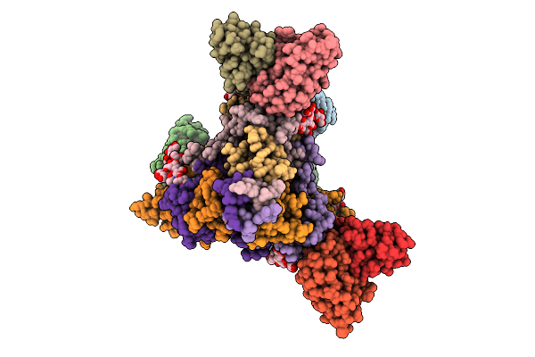 |
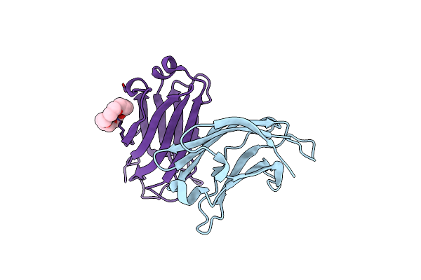
A Broadly-Neutralizing Antibody Against Ebolavirus Glycoprotein That Can Potentiate The Breadth And Neutralization Potency Of Other Anti-Glycoprotein Antibodies
Organism: Oryctolagus cuniculus, Ebolavirus
Method: ELECTRON MICROSCOPY Release Date: 2025-11-05 Classification: VIRAL PROTEIN/IMMUNE SYSTEM Ligands: NAG |
 |
Sars Cov-2 Spike Protein Ectodomain With Internal Tag, All Rbd-Down Conformation
Organism: Severe acute respiratory syndrome coronavirus 2
Method: ELECTRON MICROSCOPY Release Date: 2025-03-05 Classification: VIRAL PROTEIN Ligands: NAG |
 |
Sars Cov-2 Spike Protein Ectodomain With Internal Tag, 1Rbd-Up Conformation
Organism: Severe acute respiratory syndrome coronavirus 2
Method: ELECTRON MICROSCOPY Release Date: 2025-03-05 Classification: VIRAL PROTEIN Ligands: NAG |
 |
Cryo-Em Structure Of Sars-Cov-2 Spike Protein Ecto-Domain With Internal Tag, 1Up Rbd Conformation
Organism: Severe acute respiratory syndrome coronavirus 2
Method: ELECTRON MICROSCOPY Release Date: 2025-03-05 Classification: VIRAL PROTEIN Ligands: NAG |
 |
Cryo-Em Structure Of Sars-Cov-2 Spike Protein Ecto-Domain With Internal Tag, All Rbd Down Conformation, State-3
Organism: Severe acute respiratory syndrome coronavirus 2
Method: ELECTRON MICROSCOPY Release Date: 2025-03-05 Classification: VIRAL PROTEIN Ligands: NAG |
 |
Cryo-Em Structure Of Sars-Cov-2 Spike Protein Ecto-Domain With Internal Tag, 1Rbd Up, State-2
Organism: Severe acute respiratory syndrome coronavirus 2
Method: ELECTRON MICROSCOPY Release Date: 2025-03-05 Classification: VIRAL PROTEIN Ligands: NAG |
 |
Sars Cov-2 Spike Protein Ectodomain With Internal Tag, All Rbd-Down Conformation -C1
Organism: Severe acute respiratory syndrome coronavirus 2
Method: ELECTRON MICROSCOPY Release Date: 2025-03-05 Classification: VIRAL PROTEIN Ligands: NAG |
 |
Cryo-Em Structure Of Locally Refined Up Conformation Of Sars-Cov-2 Spike Protein Receptor Binding Domain
Organism: Severe acute respiratory syndrome coronavirus
Method: ELECTRON MICROSCOPY Release Date: 2025-03-05 Classification: VIRAL PROTEIN |
 |
Organism: Homo sapiens, Vicugna pacos
Method: X-RAY DIFFRACTION Release Date: 2025-02-26 Classification: PROTEIN TRANSPORT |
 |
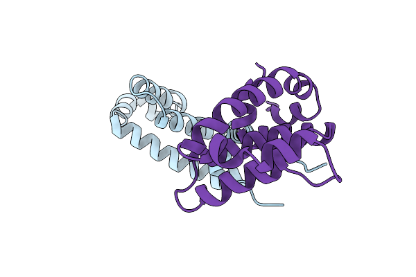
Crystal Structure Of The Human Cavin1 Hr1 Ts/Dd Mutant Domain Bound To Nanobody B7
Organism: Homo sapiens, Vicugna pacos
Method: X-RAY DIFFRACTION Release Date: 2025-02-26 Classification: PROTEIN TRANSPORT |
 |
Crystal Structure Of The Human Cavin1 Hr1 Ht/Ii Mutant Domain Bound To Nanobody B7
Organism: Homo sapiens, Vicugna pacos
Method: X-RAY DIFFRACTION Release Date: 2024-12-04 Classification: PROTEIN TRANSPORT |
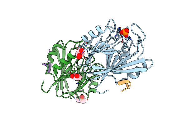 |
Crystal Structure Of Vps29 In Complex With Chaetomium Thermophilum Vps5 (71 To 80)
Organism: Mus musculus, Chaetomium thermophilum (strain dsm 1495 / cbs 144.50 / imi 039719)
Method: X-RAY DIFFRACTION Resolution:1.68 Å Release Date: 2024-07-24 Classification: PROTEIN TRANSPORT Ligands: PO4, GOL, DMS |
 |
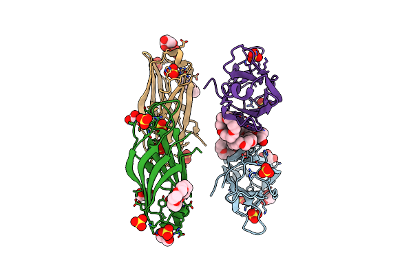
Structure Of The Inmazeb Cocktail And Resistance To Escape Against Ebola Virus
Organism: Mus musculus, Ebola virus - mayinga, zaire, 1976
Method: ELECTRON MICROSCOPY Release Date: 2023-01-25 Classification: IMMUNE SYSTEM/VIRAL PROTEIN Ligands: NAG |
 |
Organism: Homo sapiens
Method: SOLUTION NMR Release Date: 2022-08-31 Classification: STRUCTURAL PROTEIN |
 |
Crystal Structure Of The Human Snx25 Regulator Of G-Protein Signalling (Rgs) Domain
Organism: Homo sapiens
Method: X-RAY DIFFRACTION Resolution:2.40 Å Release Date: 2021-11-17 Classification: SIGNALING PROTEIN |
 |
Crystal Structure Of The Human Snx25 Regulator Of G-Protein Signalling (Rgs) Domain
Organism: Homo sapiens
Method: X-RAY DIFFRACTION Resolution:2.42 Å Release Date: 2021-11-17 Classification: SIGNALING PROTEIN Ligands: ZN, LEU, ACT |
 |
Organism: Homo sapiens
Method: X-RAY DIFFRACTION Resolution:1.68 Å Release Date: 2018-10-17 Classification: TRANSFERASE Ligands: O4B |
 |
Organism: Homo sapiens
Method: X-RAY DIFFRACTION Resolution:1.85 Å Release Date: 2018-10-17 Classification: TRANSFERASE Ligands: SO4, O4B, GOL |
 |
Organism: Homo sapiens
Method: X-RAY DIFFRACTION Resolution:2.43 Å Release Date: 2018-10-17 Classification: TRANSFERASE Ligands: FMT, IHP, O4B |
 |
Organism: Homo sapiens
Method: X-RAY DIFFRACTION Resolution:2.60 Å Release Date: 2018-10-17 Classification: TRANSFERASE Ligands: SO4, GOL |