
Search Count: 198
 |
Organism: Photorhabdus thracensis, Escherichia coli
Method: ELECTRON MICROSCOPY Release Date: 2025-03-26 Classification: DNA BINDING PROTEIN Ligands: MG, ATP |
 |
Organism: Photorhabdus thracensis, Escherichia coli
Method: ELECTRON MICROSCOPY Release Date: 2025-03-26 Classification: DNA BINDING PROTEIN Ligands: MG, ATP |
 |
Organism: Photorhabdus thracensis, Escherichia coli
Method: ELECTRON MICROSCOPY Release Date: 2025-03-26 Classification: DNA BINDING PROTEIN Ligands: MG, ATP |
 |
Organism: Photorhabdus thracensis, Escherichia coli
Method: ELECTRON MICROSCOPY Release Date: 2025-03-26 Classification: DNA BINDING PROTEIN Ligands: MG, ATP |
 |
Organism: Photorhabdus thracensis, Escherichia coli
Method: ELECTRON MICROSCOPY Release Date: 2025-03-26 Classification: DNA BINDING PROTEIN Ligands: MG, ATP |
 |
Organism: Escherichia coli, Escherichia phage t7
Method: ELECTRON MICROSCOPY Release Date: 2025-03-26 Classification: DNA BINDING PROTEIN |
 |
Organism: Escherichia coli, Escherichia phage t7
Method: ELECTRON MICROSCOPY Release Date: 2025-03-26 Classification: DNA BINDING PROTEIN |
 |
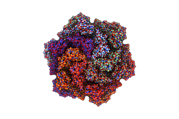
In Situ Structure Of The Nitrosopumilus Maritimus S-Layer - Six-Fold Symmetry (C6)
Organism: Nitrosopumilus maritimus scm1
Method: ELECTRON MICROSCOPY Release Date: 2024-04-17 Classification: STRUCTURAL PROTEIN |
 |
In Situ Structure Of The Nitrosopumilus Maritimus S-Layer - Composite Map Between C2 And C6
Organism: Nitrosopumilus maritimus scm1
Method: ELECTRON MICROSCOPY Release Date: 2024-04-17 Classification: STRUCTURAL PROTEIN |
 |
In Vitro Structure Of The Nitrosopumilus Maritimus S-Layer - Six-Fold Symmetry (C6)
Organism: Nitrosopumilus maritimus scm1
Method: ELECTRON MICROSCOPY Resolution:2.87 Å Release Date: 2024-04-10 Classification: STRUCTURAL PROTEIN |
 |
In Vitro Structure Of The Nitrosopumilus Maritimus S-Layer - Two-Fold Symmetry (C2)
Organism: Nitrosopumilus maritimus scm1
Method: ELECTRON MICROSCOPY Resolution:2.71 Å Release Date: 2024-04-10 Classification: STRUCTURAL PROTEIN |
 |
In Vitro Structure Of The Nitrosopumilus Maritimus S-Layer - Composite Map Between Two And Six-Fold Symmetrised
Organism: Nitrosopumilus maritimus scm1
Method: ELECTRON MICROSCOPY Resolution:2.87 Å Release Date: 2024-04-10 Classification: STRUCTURAL PROTEIN |
 |
In Situ Structure Of The Nitrosopumilus Maritimus S-Layer - Two-Fold Symmetry (C2)
Organism: Nitrosopumilus maritimus scm1
Method: ELECTRON MICROSCOPY Release Date: 2024-04-10 Classification: STRUCTURAL PROTEIN |
 |
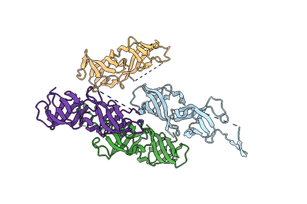
Crystal Structure Of The Hetero-Dimeric Complex From Archaeoglobus Fulgidus Prc1 And Prc2 Domains
Organism: Archaeoglobus fulgidus
Method: X-RAY DIFFRACTION Resolution:2.29 Å Release Date: 2024-04-10 Classification: CELL CYCLE |
 |
Crystal Structure Of Heterodimeric Complex Of Cdpb1 And Cdpb2 From A. Fulgidus
Organism: Archaeoglobus fulgidus
Method: X-RAY DIFFRACTION Resolution:2.29 Å Release Date: 2024-04-03 Classification: CELL CYCLE |
 |
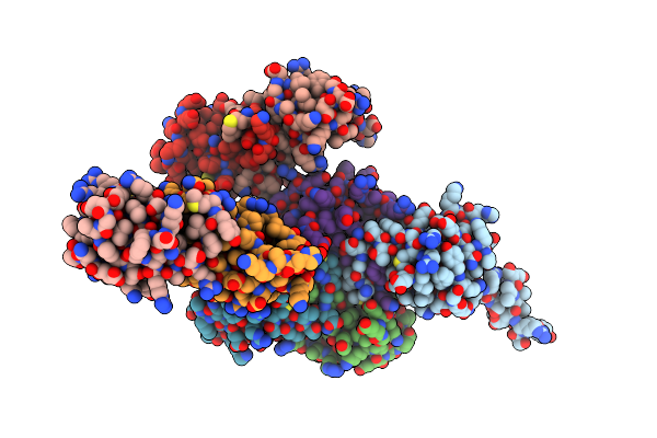
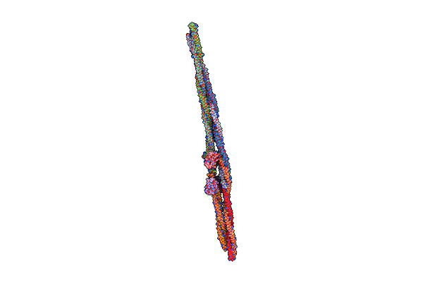
Cryo-Em Structure Of Crescentin Filaments (Stutter Mutant, C1 Symmetry And Small Box)
Organism: Caulobacter vibrioides, Camelidae
Method: ELECTRON MICROSCOPY Release Date: 2023-08-16 Classification: STRUCTURAL PROTEIN |
 |
Cryo-Em Structure Of Crescentin Filaments (Stutter Mutant, C2, Symmetry And Small Box)
Organism: Caulobacter vibrioides, Camelidae
Method: ELECTRON MICROSCOPY Release Date: 2023-08-16 Classification: STRUCTURAL PROTEIN |
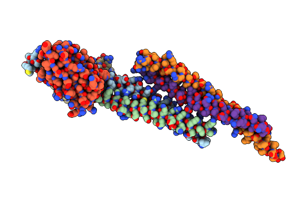 |
Cryo-Em Structure Of Crescentin Filaments (Wildtype, C1 Symmetry And Small Box)
Organism: Caulobacter vibrioides, Camelidae
Method: ELECTRON MICROSCOPY Release Date: 2023-08-16 Classification: STRUCTURAL PROTEIN |
 |
Cryo-Em Structure Of Crescentin Filaments (Wildtype, C2 Symmetry And Small Box)
Organism: Caulobacter vibrioides, Camelidae
Method: ELECTRON MICROSCOPY Release Date: 2023-08-16 Classification: STRUCTURAL PROTEIN |
 |
Cryo-Em Structure Of Crescentin Filaments (Stutter Mutant, C1 Symmetry And Large Box)
Organism: Caulobacter vibrioides, Camelidae
Method: ELECTRON MICROSCOPY Resolution:4.10 Å Release Date: 2023-08-16 Classification: STRUCTURAL PROTEIN |