
Search Count: 6
 |
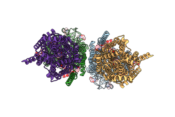
Organism: Homo sapiens
Method: ELECTRON MICROSCOPY Release Date: 2024-06-12 Classification: MEMBRANE PROTEIN Ligands: NAG |
 |
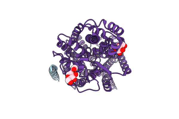
Organism: Homo sapiens
Method: ELECTRON MICROSCOPY Release Date: 2024-06-12 Classification: MEMBRANE PROTEIN Ligands: ZN, NDG, NAG |
 |
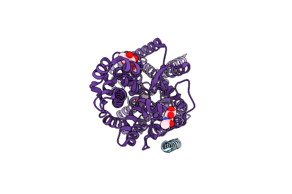
Organism: Homo sapiens
Method: ELECTRON MICROSCOPY Release Date: 2024-06-12 Classification: MEMBRANE PROTEIN Ligands: ZN, NDG, NAG |
 |
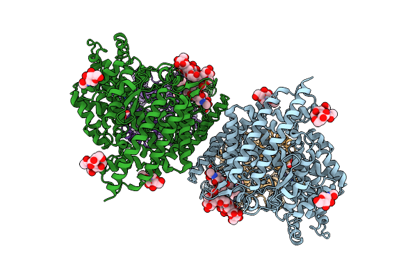
Organism: Homo sapiens
Method: ELECTRON MICROSCOPY Release Date: 2024-06-12 Classification: MEMBRANE PROTEIN Ligands: NAG, YCP, CL |
 |
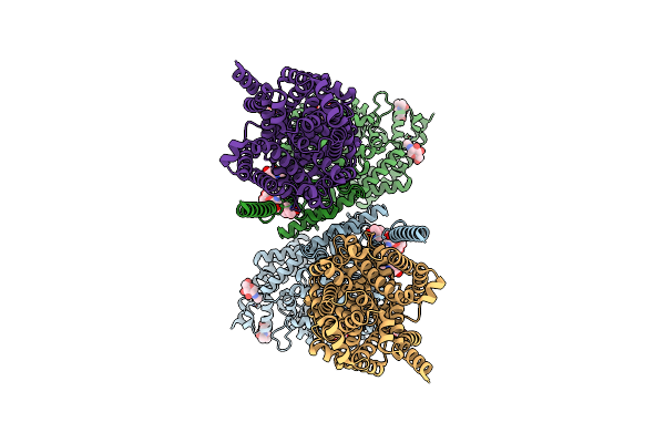
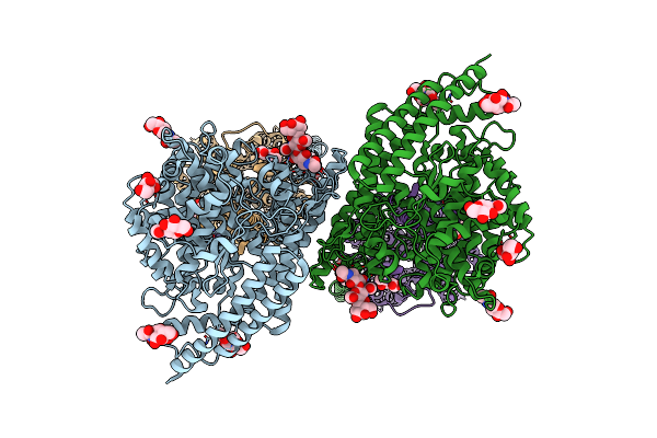
Structure Of Human Sit1:Ace2 Complex (Open Pd Conformation) Bound To L-Pipecolate
Organism: Homo sapiens
Method: ELECTRON MICROSCOPY Release Date: 2024-06-12 Classification: MEMBRANE PROTEIN Ligands: ZN, NDG, NAG, YCP, CL |
 |
Structure Of Human Sit1:Ace2 Complex (Closed Pd Conformation) Bound To L-Pipecolate
Organism: Homo sapiens
Method: ELECTRON MICROSCOPY Release Date: 2024-06-12 Classification: MEMBRANE PROTEIN Ligands: ZN, NDG, NAG, YCP |