
Search Count: 19
 |
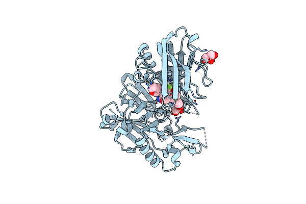
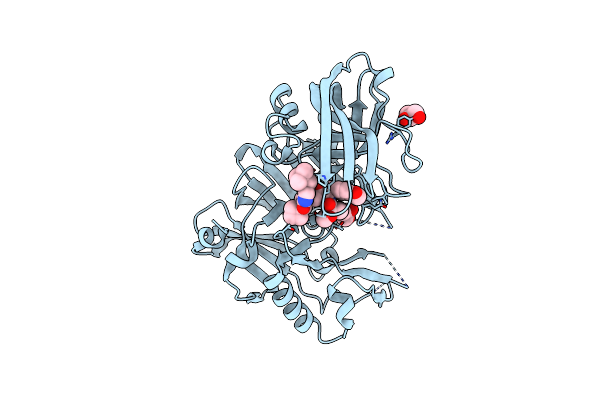
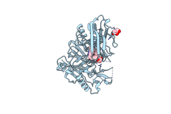
Crystal Structure Of Human Kras G12C Covalently Bound To Del Triazine Compound 5
Organism: Homo sapiens
Method: X-RAY DIFFRACTION Resolution:1.65 Å Release Date: 2025-02-26 Classification: SIGNALING PROTEIN,ONCOPROTEIN/INHIBITOR Ligands: CA, GDP, A1BH6 |
 |
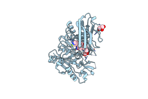
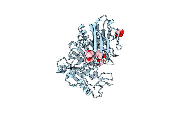
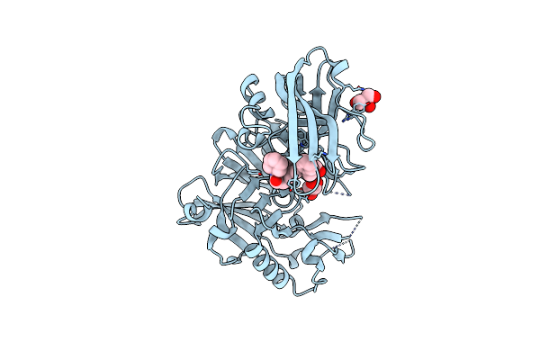
Crystal Structure Of Human Kras G12C Covalently Bound To Nopinone-Derived Naphthol Compound 21
Organism: Homo sapiens
Method: X-RAY DIFFRACTION Resolution:1.18 Å Release Date: 2025-02-26 Classification: SIGNALING PROTEIN,ONCOPROTEIN/INHIBITOR Ligands: MG, GDP, A1BH5, EDO |
 |
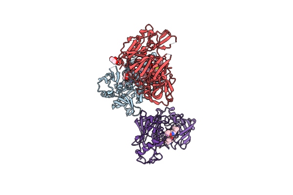
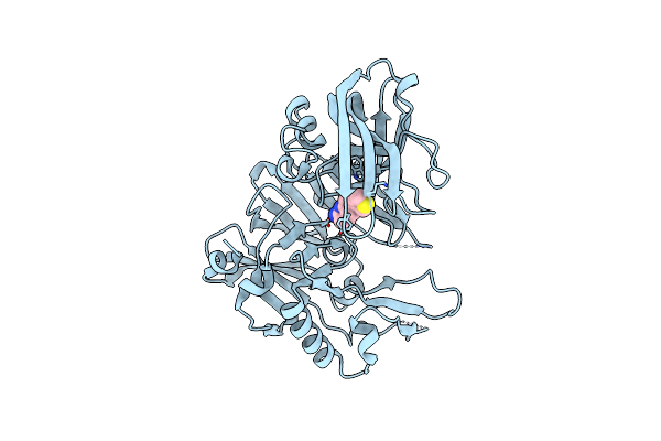
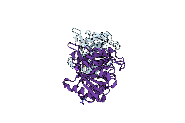
Amg 193, A Clinical Stage Mta-Cooperative Prmt5 Inhibitor, Drives Anti-Tumor Activity Preclinically And In Patients With Mtap-Deleted Cancers
Organism: Homo sapiens
Method: X-RAY DIFFRACTION Resolution:2.85 Å Release Date: 2024-10-02 Classification: TRANSFERASE/TRANSFERASE INHIBITOR Ligands: GOL, DMS, A1ATH, MTA |
 |
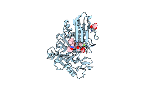
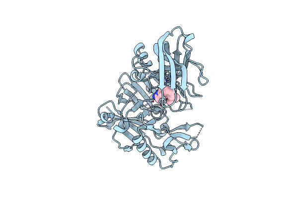
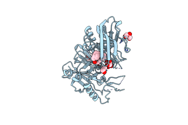
Crystal Structure Of Human Kras G12C Covalently Bound To A Phthalazine Inhibitor
Organism: Homo sapiens
Method: X-RAY DIFFRACTION Resolution:1.60 Å Release Date: 2019-12-25 Classification: SIGNALING PROTEIN/Inhibitor Ligands: MG, GDP, OJ1 |
 |
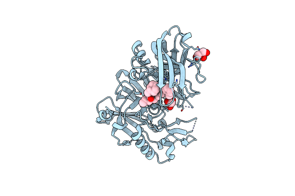
Crystal Structure Of Human Kras G12C Covalently Bound To A Quinazolinone Inhibitor
Organism: Homo sapiens
Method: X-RAY DIFFRACTION Resolution:1.50 Å Release Date: 2019-12-25 Classification: SIGNALING PROTEIN/Inhibitor Ligands: GDP, CA, OHY |
 |
Crystal Structure Of Bace1 In Complex With 2-Aminooxazoline 3-Aza-4-Fluoro-Xanthene Inhibitor 22
Organism: Homo sapiens
Method: X-RAY DIFFRACTION Resolution:1.85 Å Release Date: 2015-03-04 Classification: Hydrolase/Hydrolase Inhibitor Ligands: IOD, GOL, 3UT |
 |
Crystal Structure Of Bace1 In Complex With 2-Aminooxazoline 3-Azaxanthene Inhibitor 28
Organism: Homo sapiens
Method: X-RAY DIFFRACTION Resolution:1.80 Å Release Date: 2015-02-04 Classification: Hydrolase/hydrolase inhibitor Ligands: IOD, 43K, GOL |
 |
Organism: Homo sapiens
Method: X-RAY DIFFRACTION Resolution:2.00 Å Release Date: 2012-10-10 Classification: HYDROLASE/HYDROLASE INHIBITOR Ligands: 0K9, GOL |
 |
Structure Of Bace-1 (Beta-Secretase) In Complex With N-((2S,3R)-1-(4-Fluorophenyl)-3-Hydroxy-4-((6'-Neopentyl-3',4'-Dihydrospiro[Cyclobutane-1,2'-Pyrano[2,3-B]Pyridin]-4'-Yl)Amino)Butan-2-Yl)Acetamide
Organism: Homo sapiens
Method: X-RAY DIFFRACTION Resolution:2.50 Å Release Date: 2012-10-10 Classification: HYDROLASE/Hydrolase Inhibitor Ligands: IOD, GOL, 0MP |
 |
Structure Of Bace Bound To (S)-4-(3'-Methoxy-[1,1'-Biphenyl]-3-Yl)-1,4-Dimethyl-6-Oxotetrahydropyrimidin-2(1H)-Iminium
Organism: Homo sapiens
Method: X-RAY DIFFRACTION Resolution:1.74 Å Release Date: 2012-10-10 Classification: HYDROLASE/HYDROLASE INHIBITOR Ligands: H24, TLA |
 |
Structure Of Bace-1 (Beta-Secretase) In Complex With (2R)-N-((2S,3R)-1-(Benzo[D][1,3]Dioxol-5-Yl)-3-Hydroxy-4-((S)-6'-Neopentyl-3',4'-Dihydrospiro[Cyclobutane-1,2'-Pyrano[2,3-B]Pyridine]-4'-Ylamino)Butan-2-Yl)-2-Methoxypropanamide
Organism: Homo sapiens
Method: X-RAY DIFFRACTION Resolution:2.50 Å Release Date: 2012-04-18 Classification: HYDROLASE/HYDROLASE INHIBITOR Ligands: IOD, GOL, 0KN |
 |
Structure Of Bace-1 (Beta-Secretase) In Complex With (R)-3-(2-Amino-6-O-Tolylquinolin-3-Yl)-N-((R)-2,2-Dimethyltetrahydro-2H-Pyran-4-Yl)-2-Methylpropanamide
Organism: Homo sapiens
Method: X-RAY DIFFRACTION Resolution:2.50 Å Release Date: 2011-08-31 Classification: Hydrolase/Hydrolase Inhibitor Ligands: IOD, GOL, 3RS |
 |
Structure Of Bace-1 (Beta-Secretase) In Complex With 6-(Thiophen-3-Yl)Quinolin-2-Amine
Organism: Homo sapiens
Method: X-RAY DIFFRACTION Resolution:2.48 Å Release Date: 2011-08-31 Classification: Hydrolase/Hydrolase Inhibitor Ligands: IOD, RSV |
 |
Structure Of Bace-1 (Beta-Secretase) In Complex With 6-(2-(3,3-Dimethylbut-1-Ynyl)Phenyl)Quinolin-2-Amine
Organism: Homo sapiens
Method: X-RAY DIFFRACTION Resolution:2.70 Å Release Date: 2011-08-31 Classification: Hydrolase/Hydrolase Inhibitor Ligands: IOD, RTH |
 |
Structure Of Bace-1 (Beta-Secretase) In Complex With 3-(2-Aminoquinolin-3-Yl)-N-Cyclohexyl-N-Methylpropanamide
Organism: Homo sapiens
Method: X-RAY DIFFRACTION Resolution:2.76 Å Release Date: 2011-08-31 Classification: Hydrolase/Hydrolase Inhibitor Ligands: IOD, GOL, RTM |
 |
Structure Of Bace-1 (Beta-Secretase) In Complex With 3-(2-Amino-6-O-Tolylquinolin-3-Yl)-N-Cyclohexylpropanamide
Organism: Homo sapiens
Method: X-RAY DIFFRACTION Resolution:2.70 Å Release Date: 2011-08-31 Classification: Hydrolase/Hydrolase Inhibitor Ligands: IOD, GOL, RTN |
 |
Structure Of Bace-1 (Beta-Secretase) In Complex With 3-(2-Aminoquinolin-3-Yl)-N-(Cyclohexylmethyl)Propanamide
Organism: Homo sapiens
Method: X-RAY DIFFRACTION Resolution:2.30 Å Release Date: 2011-08-31 Classification: Hydrolase/Hydrolase Inhibitor Ligands: IOD, GOL, 3RU |
 |
Structure Of Bace-1 (Beta-Secretase) In Complex With 2-((2-Amino-6-O-Tolylquinolin-3-Yl)Methyl)-N-(Cyclohexylmethyl)Pentanamide
Organism: Homo sapiens
Method: X-RAY DIFFRACTION Resolution:2.65 Å Release Date: 2011-08-31 Classification: Hydrolase/Hydrolase Inhibitor Ligands: IOD, GOL, RVI |
 |
Three Dimensional Structure And Catalytic Mechanism Of 6- Phosphogluconolactonase From Trypanosoma Brucei
Organism: Trypanosoma brucei
Method: X-RAY DIFFRACTION Resolution:2.10 Å Release Date: 2007-01-03 Classification: HYDROLASE Ligands: ZN, HG, K |