
Search Count: 76
 |
Organism: Homo sapiens
Method: ELECTRON MICROSCOPY Release Date: 2025-10-08 Classification: PROTEIN FIBRIL |
 |
Organism: Corynebacterium diphtheriae nctc 13129
Method: X-RAY DIFFRACTION Release Date: 2025-09-03 Classification: TRANSPORT PROTEIN Ligands: HEM, PEG, EDO, GOL |
 |
Organism: Corynebacterium diphtheriae nctc 13129
Method: X-RAY DIFFRACTION Release Date: 2025-09-03 Classification: TRANSPORT PROTEIN Ligands: HEM, GOL, PEG, EDO, SO4 |
 |
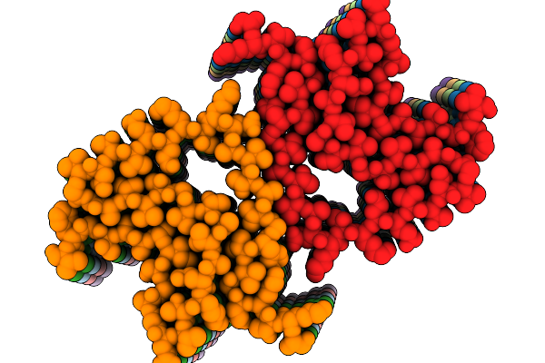
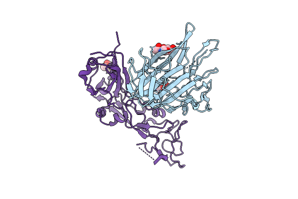
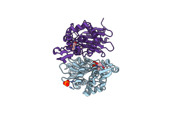
Crystal Structure Of The Porcine Hemagglutinating Encephalomyelitis Virus (Phev) Receptor Binding Domain In Complex With Porcine Dpep1.
Organism: Sus scrofa, Porcine hemagglutinating encephalomyelitis virus
Method: X-RAY DIFFRACTION Release Date: 2025-08-06 Classification: VIRAL PROTEIN Ligands: ZN, NAG |
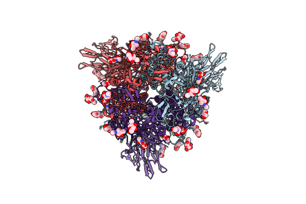 |
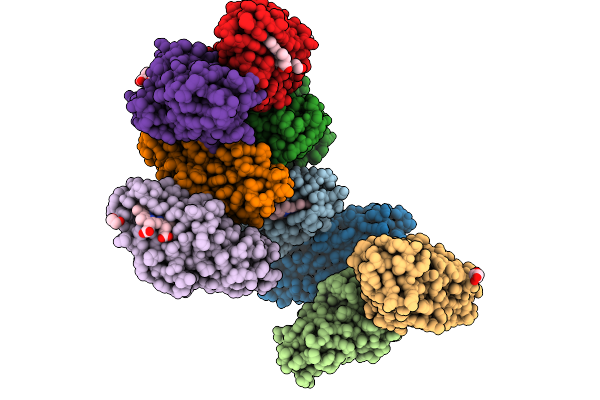
Porcine Hemagglutinating Encephalomyelitis Virus (Phev) Spike In The Closed Conformation, Apo State
Organism: Porcine hemagglutinating encephalomyelitis virus
Method: ELECTRON MICROSCOPY Release Date: 2025-08-06 Classification: VIRAL PROTEIN Ligands: NAG |
 |
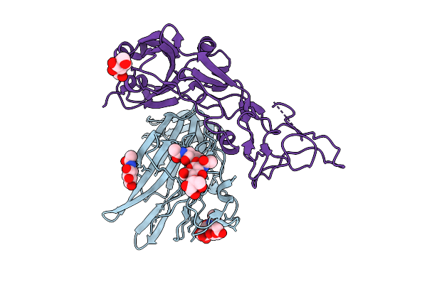
Local Refinement Of The N-Terminal Domain (Ntd) And Receptor Binding Domain (Rbd) From The Porcine Hemagglutinating Encephalomyelitis Virus (Phev) Spike In The Closed Conformation
Organism: Porcine hemagglutinating encephalomyelitis virus
Method: ELECTRON MICROSCOPY Release Date: 2025-08-06 Classification: VIRAL PROTEIN Ligands: NAG |
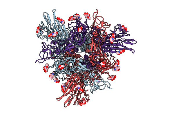 |
Porcine Hemagglutinating Encephalomyelitis Virus (Phev) Spike Protein In The Closed Conformation Bound To 9-O-Ac-Sia.
Organism: Porcine hemagglutinating encephalomyelitis virus
Method: ELECTRON MICROSCOPY Release Date: 2025-08-06 Classification: VIRAL PROTEIN Ligands: NAG, 5N6 |
 |
Local Refinement Of The N-Terminal Domain (Ntd) And Receptor Binding Domain (Rbd) From The Porcine Hemagglutinating Encephalomyelitis Virus (Phev) Spike In The Closed Conformation Bound To 9-O-Ac-Sia
Organism: Porcine hemagglutinating encephalomyelitis virus
Method: ELECTRON MICROSCOPY Release Date: 2025-08-06 Classification: VIRAL PROTEIN Ligands: NAG, 5N6 |
 |
Local Refinement Of The N-Terminal Domain (Ntd) From The Porcine Hemagglutinating Encephalomyelitis Virus (Phev) Spike In The Open Conformation Bound To 9-O-Ac-Sia
Organism: Porcine hemagglutinating encephalomyelitis virus
Method: ELECTRON MICROSCOPY Release Date: 2025-08-06 Classification: VIRAL PROTEIN Ligands: NAG, 5N6 |
 |
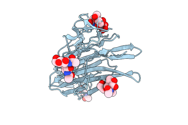
Organism: Carica papaya
Method: X-RAY DIFFRACTION Release Date: 2025-06-18 Classification: HYDROLASE |
 |
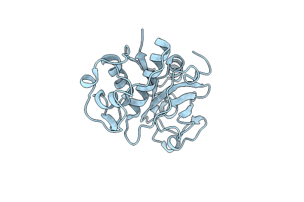
Organism: Escherichia coli
Method: X-RAY DIFFRACTION Release Date: 2025-06-18 Classification: HYDROLASE Ligands: PO4, K |
 |
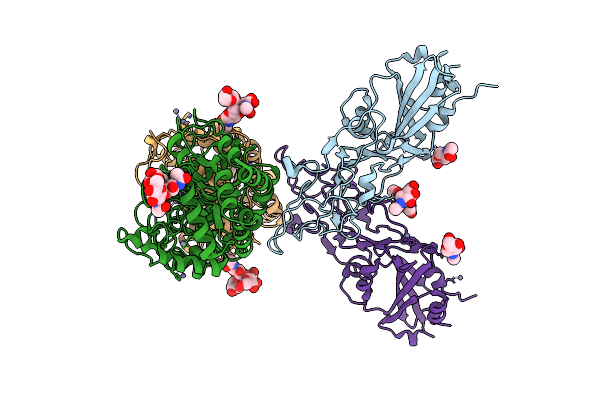
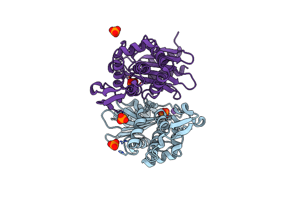
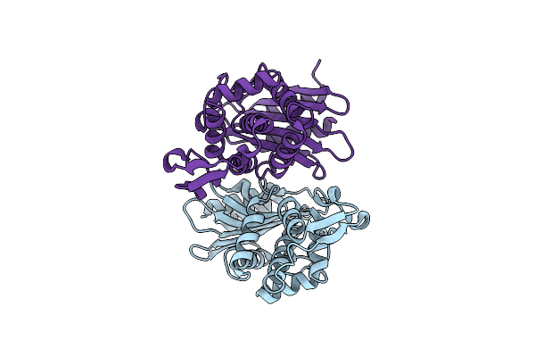
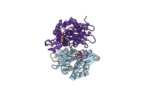
X-Ray Diffraction Structure Of Ctx-M-14 Beta-Lactamase Co-Crystallized With Avibactam
Organism: Escherichia coli
Method: X-RAY DIFFRACTION Release Date: 2025-06-18 Classification: HYDROLASE Ligands: NXL, GOL, PO4 |
 |
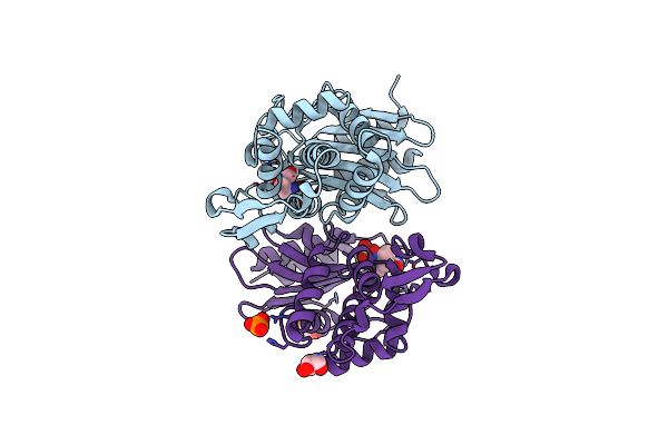
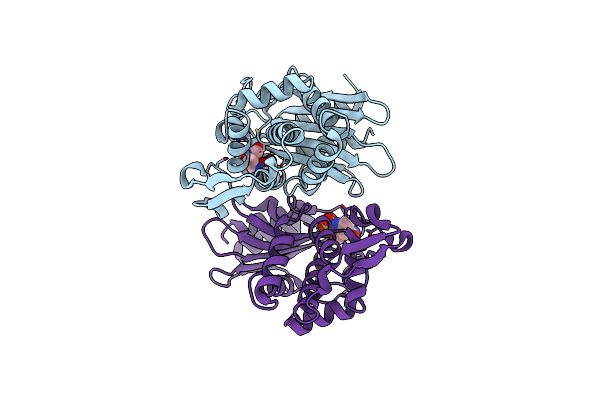
X-Ray Diffraction Structure Of Ctx-M-14 Beta-Lactamase Soaked With Avibactam
Organism: Escherichia coli
Method: X-RAY DIFFRACTION Release Date: 2025-06-18 Classification: HYDROLASE Ligands: NXL, PO4 |
 |
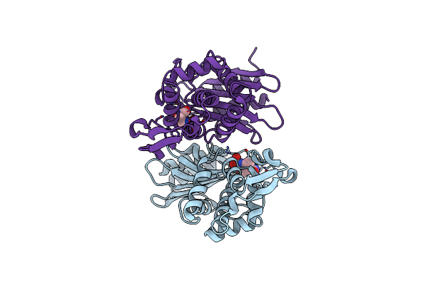
X-Ray Diffraction Structure Of The Ctx-M-14 Beta-Lactamase-Avibactam Complex An Inhibitor Cocktail-Soaked Crystal
Organism: Escherichia coli
Method: X-RAY DIFFRACTION Release Date: 2025-06-18 Classification: HYDROLASE Ligands: NXL, PO4 |
 |
Organism: Escherichia coli
Method: ELECTRON CRYSTALLOGRAPHY Release Date: 2025-06-18 Classification: HYDROLASE |
 |
Microed Structure Of The Ctx-M-14 Beta-Lactamase-Avibactam Complex From Inhibitor Cocktail-Soaked Crystals
Organism: Escherichia coli
Method: ELECTRON CRYSTALLOGRAPHY Release Date: 2025-06-18 Classification: HYDROLASE Ligands: NXL |
 |
Organism: Escherichia coli
Method: ELECTRON CRYSTALLOGRAPHY Release Date: 2025-06-18 Classification: HYDROLASE Ligands: NXL |
 |
Microed Structure Of Ctx-M-14 Beta-Lactamase Co-Crystallized With Avibactam
Organism: Escherichia coli
Method: ELECTRON CRYSTALLOGRAPHY Release Date: 2025-06-18 Classification: HYDROLASE Ligands: NXL |
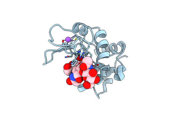 |
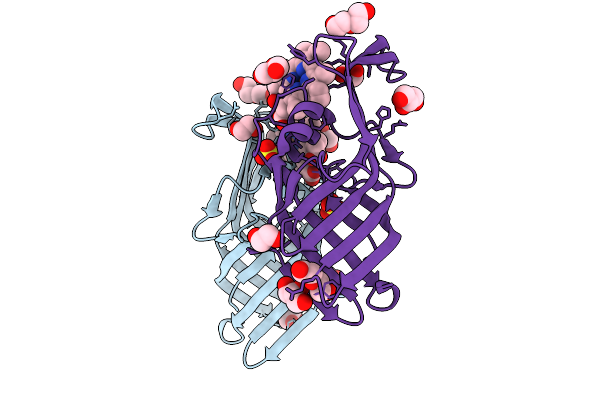
X-Ray Diffraction Structure Of Lysozyme Co-Crystallized With N,N',N"-Triacetylchitotriose
Organism: Gallus gallus
Method: X-RAY DIFFRACTION Release Date: 2025-06-18 Classification: HYDROLASE Ligands: CL, NA |
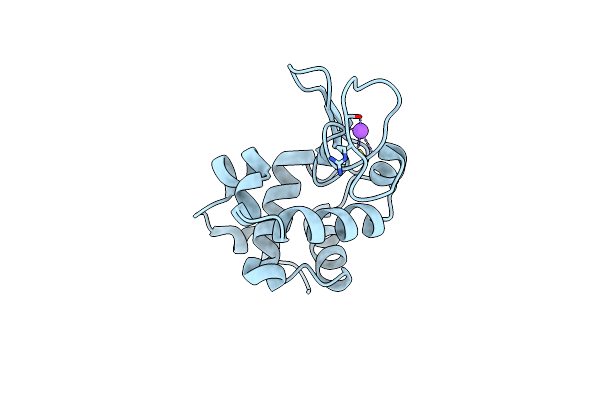 |
Organism: Gallus gallus
Method: X-RAY DIFFRACTION Release Date: 2025-06-18 Classification: HYDROLASE Ligands: CL, NA |