
Search Count: 20
 |
Organism: Lampyridae, Oryctolagus cuniculus, Bos taurus
Method: ELECTRON MICROSCOPY Release Date: 2025-07-30 Classification: RIBOSOME/ANTIBIOTIC Ligands: MG, ZN, 5GP, ANM, SPD, SPM, K, GTP, SER |
 |
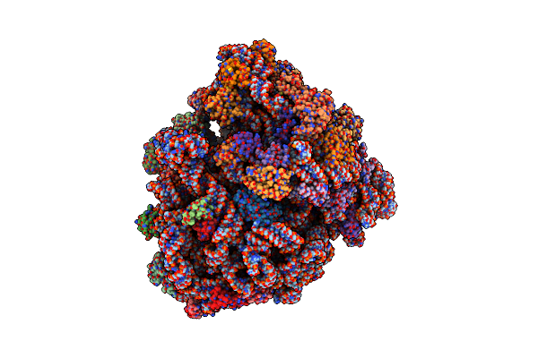
Codon Sampling State Of Elongation Inhibitor-Treated Mammalian Ribosomes Obtained From Merged Datasets
Organism: Lampyridae, Oryctolagus cuniculus, Bos taurus
Method: ELECTRON MICROSCOPY Release Date: 2025-07-30 Classification: RIBOSOME/ANTIBIOTIC Ligands: MG, ZN, 5GP, ANM, SPD, K, GTP, SER |
 |
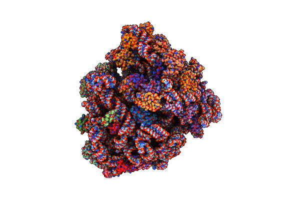
Post-Decoding/Post-Hydrolysis State Obtained From Anisomycin-Treated Mammalian Ribosomes
Organism: Lampyridae, Oryctolagus cuniculus, Bos taurus
Method: ELECTRON MICROSCOPY Release Date: 2025-07-30 Classification: RIBOSOME/ANTIBIOTIC Ligands: MG, ZN, 5GP, ANM, SPD, K, SER |
 |
Post-Decoding Post-Hydrolysis State Obtained From Merged Datasets Of Elongation Inhibitor-Treated Mammalian Ribosomes
Organism: Lampyridae, Oryctolagus cuniculus, Bos taurus
Method: ELECTRON MICROSCOPY Release Date: 2025-07-30 Classification: RIBOSOME/ANTIBIOTIC Ligands: MG, ZN, 5GP, ANM, SPD, K, SER |
 |
Organism: Oryctolagus cuniculus
Method: ELECTRON MICROSCOPY Release Date: 2025-07-30 Classification: RIBOSOME/ANTIBIOTIC Ligands: MG, ANM, SPD, ZN, GDP |
 |
Anisomycin-Bound Mammalian Ribosome With Partially Accommodated A-Site Trna
Organism: Lampyridae, Oryctolagus cuniculus, Bos taurus
Method: ELECTRON MICROSCOPY Release Date: 2025-07-30 Classification: RIBOSOME/ANTIBIOTIC Ligands: MG, ZN, ANM, SPD, SER |
 |
Organism: Oryctolagus cuniculus
Method: ELECTRON MICROSCOPY Release Date: 2025-07-30 Classification: RIBOSOME/ANTIBIOTIC Ligands: MG, ZN, GDP |
 |
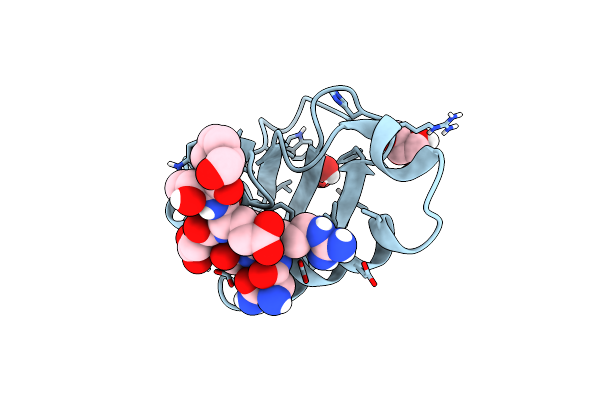
Organism: Homo sapiens
Method: X-RAY DIFFRACTION Resolution:1.80 Å Release Date: 2022-07-27 Classification: PEPTIDE BINDING PROTEIN Ligands: IPA |
 |
Organism: Mus musculus
Method: ELECTRON MICROSCOPY Release Date: 2021-11-03 Classification: RIBOSOME Ligands: ZN, MG, GDP |
 |
Organism: Mus musculus
Method: ELECTRON MICROSCOPY Release Date: 2021-11-03 Classification: RIBOSOME Ligands: ZN, MG, GDP |
 |
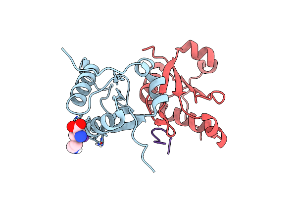
70S Ribosome Bound To Hiv Frameshifting Stem-Loop (Fss) And P/E Trna (Rotated Conformation)
Organism: Escherichia coli, Human immunodeficiency virus 1
Method: ELECTRON MICROSCOPY Release Date: 2020-06-03 Classification: RIBOSOME Ligands: MG |
 |
70S Ribosome Bound To Hiv Frameshifting Stem-Loop (Fss) And P-Site Trna (Non-Rotated Conformation, Structure I)
Organism: Human immunodeficiency virus 1, Escherichia coli
Method: ELECTRON MICROSCOPY Release Date: 2020-06-03 Classification: RIBOSOME Ligands: MG |
 |
70S Ribosome Bound To Hiv Frameshifting Stem-Loop (Fss) And P-Site Trna (Non-Rotated Conformation, Structure Ii)
Organism: Human immunodeficiency virus 1, Escherichia coli
Method: ELECTRON MICROSCOPY Release Date: 2020-06-03 Classification: RIBOSOME Ligands: MG |
 |
Organism: Mus musculus
Method: ELECTRON MICROSCOPY Release Date: 2019-03-13 Classification: PROTEIN FIBRIL |
 |
Organism: Homo sapiens
Method: ELECTRON MICROSCOPY Release Date: 2019-03-13 Classification: PROTEIN FIBRIL |
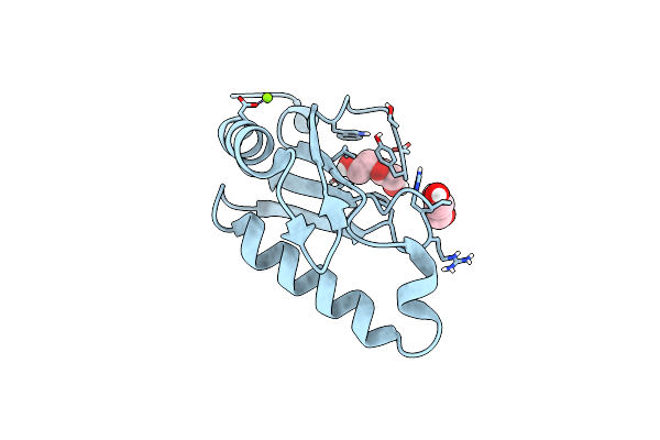 |
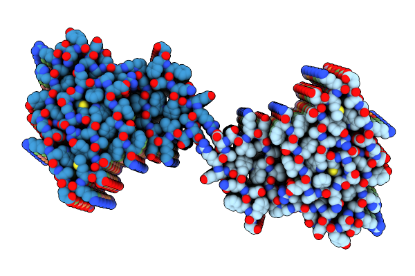
Organism: Homo sapiens
Method: X-RAY DIFFRACTION Resolution:1.13 Å Release Date: 2019-01-02 Classification: PEPTIDE BINDING PROTEIN Ligands: CL, PEG, MG, GOL |
 |
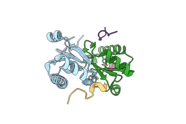
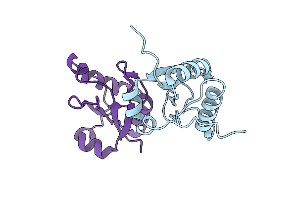
Structure Of Hiv Tat-Specific Factor 1 U2Af Homology Motif Bound To U2Af Ligand Motif 4
Organism: Homo sapiens
Method: X-RAY DIFFRACTION Resolution:1.89 Å Release Date: 2019-01-02 Classification: PROTEIN BINDING Ligands: GOL, FMT |
 |
Structure Of Hiv Tat-Specific Factor 1 U2Af Homology Motif Bound To Sf3B1 Ulm5
Organism: Homo sapiens
Method: X-RAY DIFFRACTION Resolution:2.10 Å Release Date: 2019-01-02 Classification: PEPTIDE BINDING PROTEIN Ligands: PEG, GOL |
 |
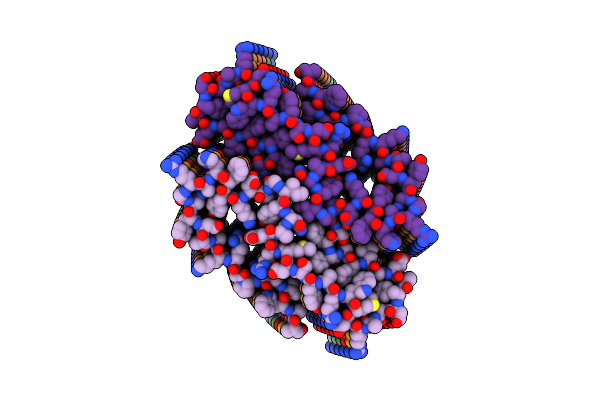
Organism: Homo sapiens
Method: X-RAY DIFFRACTION Resolution:2.20 Å Release Date: 2014-05-14 Classification: TRANSCRIPTION Ligands: CL |
 |
Organism: Homo sapiens
Method: X-RAY DIFFRACTION Resolution:1.74 Å Release Date: 2014-05-14 Classification: TRANSCRIPTION Ligands: K, CL, LYS |