
Search Count: 269
 |
Organism: Leptospirillum ferriphilum
Method: ELECTRON MICROSCOPY Release Date: 2025-12-03 Classification: DNA BINDING PROTEIN |
 |
Cryo-Em Structure Of The Type Ii Secretion System Protein From Acidithiobacillus Caldus
Organism: Acidithiobacillus caldus (strain sm-1)
Method: ELECTRON MICROSCOPY Resolution:2.39 Å Release Date: 2025-11-19 Classification: TOXIN |
 |
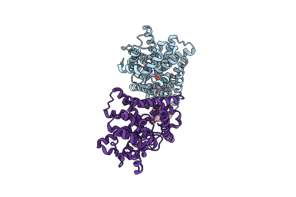
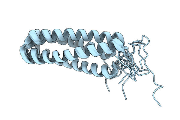
Nrbf2 Coiled Coil Domain Promotes Autophagy By Strengthening Association With Vps15 In The Pi3Kc3 Complex
Organism: Mus musculus
Method: X-RAY DIFFRACTION Resolution:2.25 Å Release Date: 2025-11-12 Classification: STRUCTURAL PROTEIN |
 |
Organism: Thermus thermophilus hb8, Discosoma sp.
Method: ELECTRON MICROSCOPY Release Date: 2025-10-29 Classification: FLUORESCENT PROTEIN Ligands: CA, 2OP |
 |
Cryo-Em Structure Of The Type Ii Secretion System Protein From Vibrio Cholerae
Organism: Vibrio cholerae
Method: ELECTRON MICROSCOPY Release Date: 2025-10-29 Classification: PROTEIN TRANSPORT |
 |
Organism: Homo sapiens
Method: X-RAY DIFFRACTION Resolution:1.85 Å Release Date: 2025-09-10 Classification: APOPTOSIS Ligands: MG, PD |
 |
Organism: Homo sapiens
Method: X-RAY DIFFRACTION Resolution:1.53 Å Release Date: 2025-09-10 Classification: APOPTOSIS |
 |
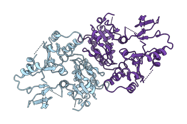
Crystal Structure Of The Periplasmic Domain Of Sensor Protein Evgs From Escherichia Coli Str. K-12 Substr. Mg1655
Organism: Escherichia coli
Method: X-RAY DIFFRACTION Resolution:2.70 Å Release Date: 2025-07-09 Classification: SIGNALING PROTEIN |
 |
Organism: Homo sapiens
Method: X-RAY DIFFRACTION Resolution:2.30 Å Release Date: 2025-06-25 Classification: HYDROLASE Ligands: ZN, MG, A1EBE |
 |
Crystal Structure Of Dna-Binding Transcriptional Activator Evga From Escherichia Coli Str. K-12 Substr. Mg1655
Organism: Escherichia coli
Method: X-RAY DIFFRACTION Resolution:2.90 Å Release Date: 2025-06-18 Classification: DNA BINDING PROTEIN |
 |
Organism: Monkeypox virus
Method: ELECTRON MICROSCOPY Release Date: 2025-06-18 Classification: VIRAL PROTEIN |
 |
Organism: Monkeypox virus
Method: ELECTRON MICROSCOPY Release Date: 2025-06-18 Classification: VIRAL PROTEIN/DNA Ligands: ADP, ATP, ZN |
 |
The Cryo-Em Structure Of Mpxv E5 In Complex With Ssdna Focused On Primase And Zn Binding Domain
Organism: Monkeypox virus
Method: ELECTRON MICROSCOPY Release Date: 2025-06-18 Classification: VIRAL PROTEIN Ligands: ZN, ATP |
 |
The Cryo-Em Structure Of Mpxv E5 In Complex With Ssdna In Intermediate State 1
Organism: Monkeypox virus
Method: ELECTRON MICROSCOPY Release Date: 2025-06-18 Classification: VIRAL PROTEIN/DNA Ligands: ATP, MG, ADP |
 |
The Cryo-Em Structure Of Mpxv E5 In Complex With Ssdna In Intermediate State 3
Organism: Monkeypox virus
Method: ELECTRON MICROSCOPY Release Date: 2025-06-18 Classification: VIRAL PROTEIN/DNA Ligands: AMP, ATP, MG, ADP |
 |
Organism: Monkeypox virus
Method: ELECTRON MICROSCOPY Release Date: 2025-06-18 Classification: VIRAL PROTEIN |
 |
Organism: Homo sapiens
Method: ELECTRON MICROSCOPY Release Date: 2025-05-28 Classification: RNA BINDING PROTEIN Ligands: ZN |
 |
Organism: Homo sapiens, Vicugna pacos, Camelus dromedarius
Method: X-RAY DIFFRACTION Resolution:3.28 Å Release Date: 2025-05-21 Classification: OXIDOREDUCTASE/IMMUNE SYSTEM Ligands: ZN |
 |
De Novo Design Of Proteins That Bind Naphthalenediimides, Powerful Photooxidants With Tunable Photophysical Properties
|
 |
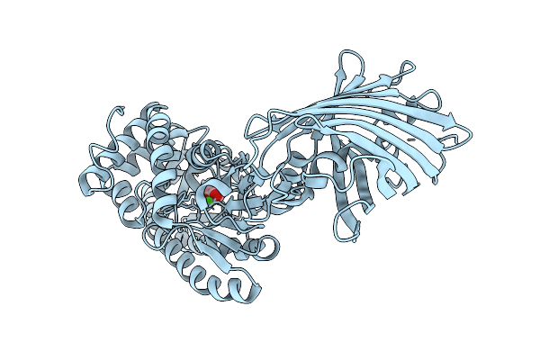
Crystal Structure Of Human Kras G12C Covalently Bound To Del Triazine Compound 5
Organism: Homo sapiens
Method: X-RAY DIFFRACTION Resolution:1.65 Å Release Date: 2025-02-26 Classification: SIGNALING PROTEIN,ONCOPROTEIN/INHIBITOR Ligands: CA, GDP, A1BH6 |