Planned Maintenance: Some services may turn out to be unavailable from 15th January, 2026 to 16th January, 2026. We apologize for the inconvenience!

Planned Maintenance: Some services may turn out to be unavailable from 15th January, 2026 to 16th January, 2026. We apologize for the inconvenience!
 |
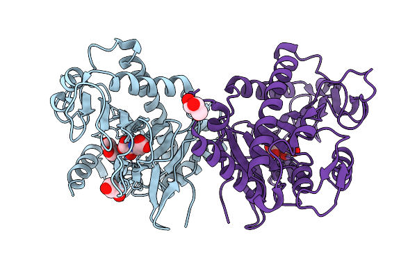
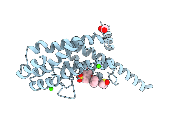
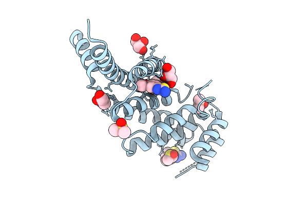
Organism: Sporormiella sp.
Method: X-RAY DIFFRACTION Release Date: 2026-01-07 Classification: OXIDOREDUCTASE Ligands: FE, CL |
 |
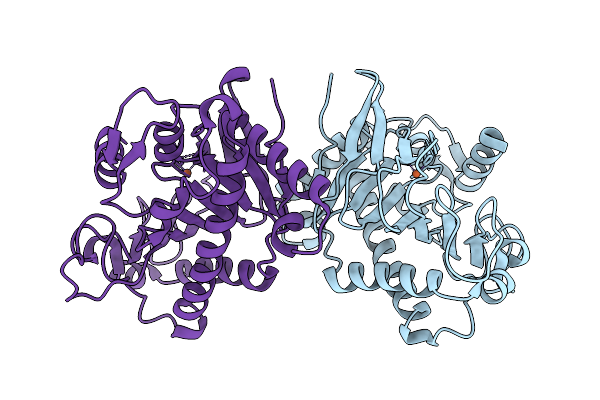
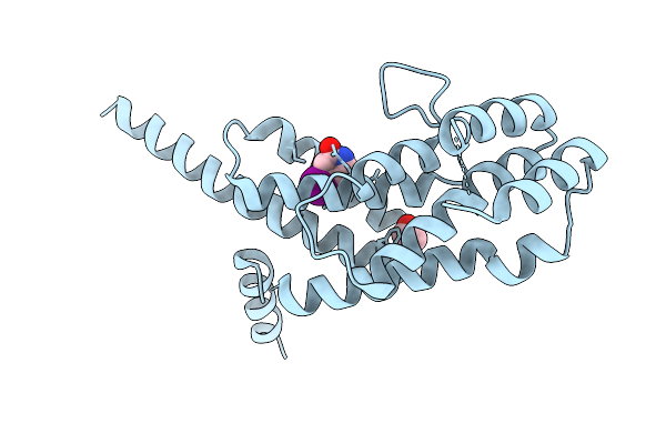
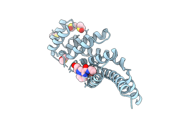
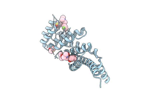
Cryo-Em Structure Of The Cytosolic Armh2-Efcab9-Catsperz Subcomplex Of The Mouse Catspermasome
Organism: Mus musculus
Method: ELECTRON MICROSCOPY Release Date: 2025-12-31 Classification: CYTOSOLIC PROTEIN |
 |
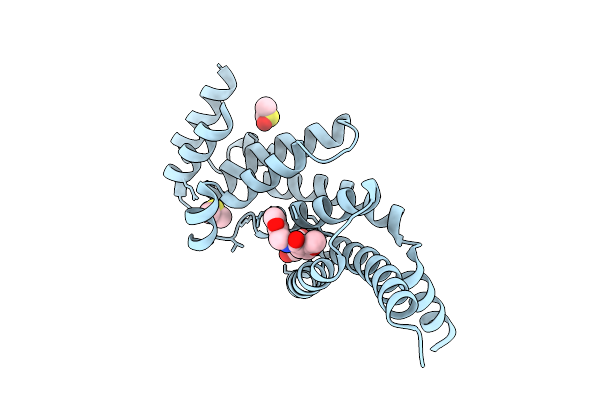
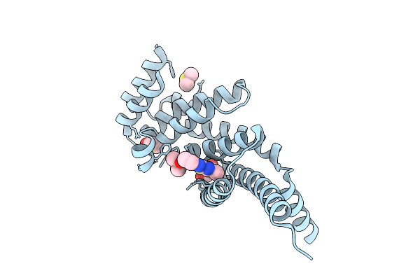
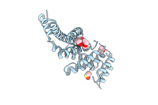
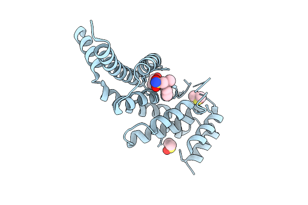
The Ring Expansion Oxygenase Spoc In Complex With Mn And N-Oxalylglycine (Nog)
Organism: Sporormiella sp.
Method: X-RAY DIFFRACTION Release Date: 2025-12-24 Classification: OXIDOREDUCTASE Ligands: MN, OGA, GOL |
 |
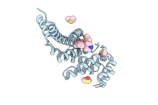
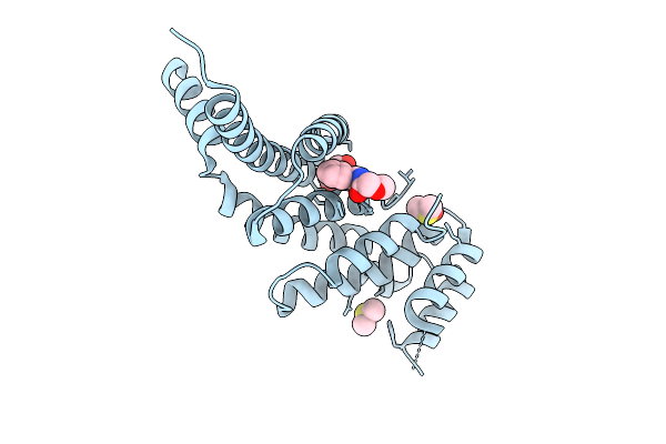
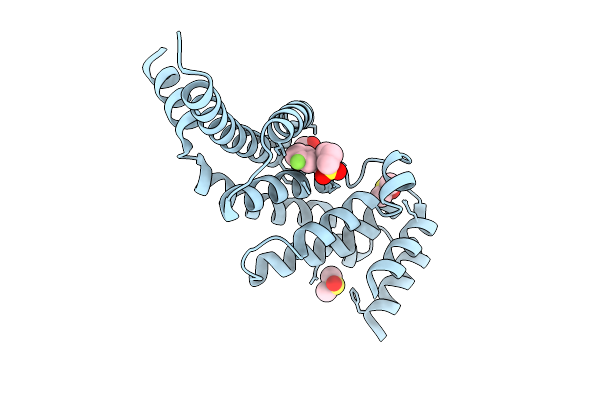
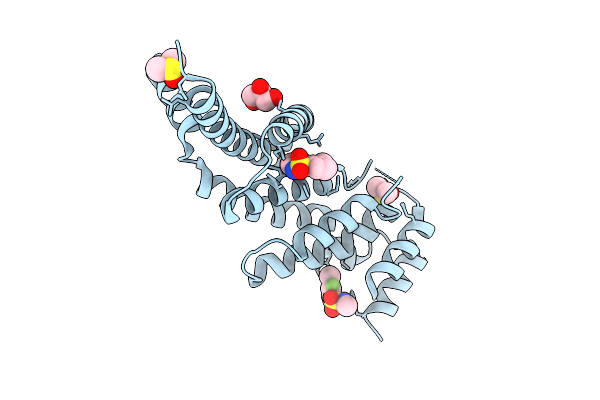
Organism: Sporormiella sp.
Method: X-RAY DIFFRACTION Release Date: 2025-12-24 Classification: OXIDOREDUCTASE Ligands: FE, CL |
 |
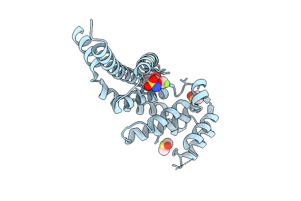
Organism: Homo sapiens
Method: X-RAY DIFFRACTION Release Date: 2025-12-03 Classification: PROTEIN BINDING Ligands: GOL, A1IU1, DMS |
 |
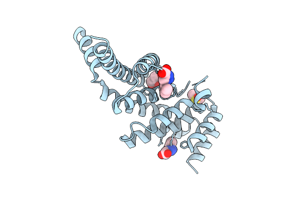
Organism: Homo sapiens
Method: X-RAY DIFFRACTION Release Date: 2025-12-03 Classification: PROTEIN BINDING Ligands: GOL, A1IV7, DMS |
 |
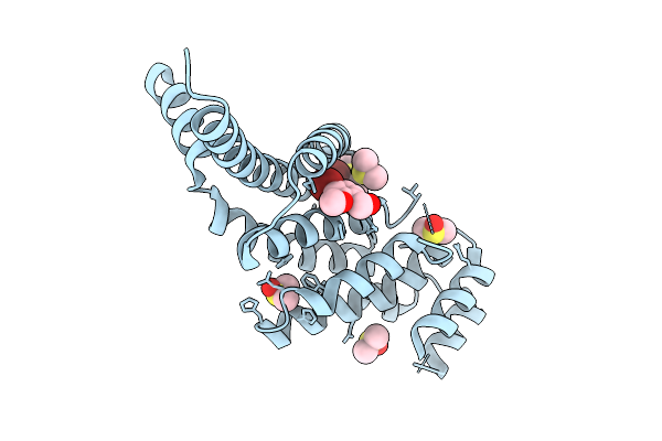
Organism: Homo sapiens
Method: X-RAY DIFFRACTION Release Date: 2025-12-03 Classification: PROTEIN BINDING Ligands: HHQ, EDO |
 |
Organism: Homo sapiens
Method: X-RAY DIFFRACTION Release Date: 2025-12-03 Classification: PROTEIN BINDING Ligands: GOL, LL0, DMS |
 |
Organism: Homo sapiens
Method: X-RAY DIFFRACTION Release Date: 2025-12-03 Classification: PROTEIN BINDING Ligands: GOL, UJK, DMS |
 |
Organism: Homo sapiens
Method: X-RAY DIFFRACTION Release Date: 2025-12-03 Classification: PROTEIN BINDING Ligands: UUG, DMS |
 |
Organism: Homo sapiens
Method: X-RAY DIFFRACTION Release Date: 2025-11-26 Classification: PROTEIN BINDING Ligands: A1IUC, DMS, EDO, CA |
 |
Organism: Homo sapiens
Method: X-RAY DIFFRACTION Release Date: 2025-11-26 Classification: PROTEIN BINDING Ligands: GOL, UUP, DMS |
 |
Organism: Homo sapiens
Method: X-RAY DIFFRACTION Release Date: 2025-11-26 Classification: PROTEIN BINDING Ligands: GOL, JFP, DMS |
 |
Organism: Homo sapiens
Method: X-RAY DIFFRACTION Release Date: 2025-11-26 Classification: PROTEIN BINDING Ligands: GOL, T9Q, DMS |
 |
Organism: Homo sapiens
Method: X-RAY DIFFRACTION Release Date: 2025-11-26 Classification: PROTEIN BINDING Ligands: NZ1, GOL, DMS |
 |
Organism: Homo sapiens
Method: X-RAY DIFFRACTION Release Date: 2025-11-26 Classification: PROTEIN BINDING Ligands: TT3, GOL, DMS |
 |
Organism: Homo sapiens
Method: X-RAY DIFFRACTION Release Date: 2025-11-26 Classification: PROTEIN BINDING Ligands: GOL, JP4, DMS |
 |
Organism: Homo sapiens
Method: X-RAY DIFFRACTION Release Date: 2025-11-26 Classification: PROTEIN BINDING Ligands: GOL, WHM, DMS |
 |
Organism: Homo sapiens
Method: X-RAY DIFFRACTION Release Date: 2025-11-26 Classification: PROTEIN BINDING Ligands: GOL, VW4, DMS |
 |
Organism: Homo sapiens
Method: X-RAY DIFFRACTION Release Date: 2025-11-26 Classification: PROTEIN BINDING Ligands: PK4, GOL, DMS |