
Search Count: 38
 |
Organism: Staphylococcus aureus
Method: ELECTRON MICROSCOPY Release Date: 2022-07-06 Classification: RIBOSOME |
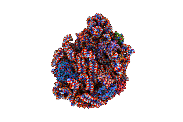 |
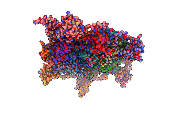
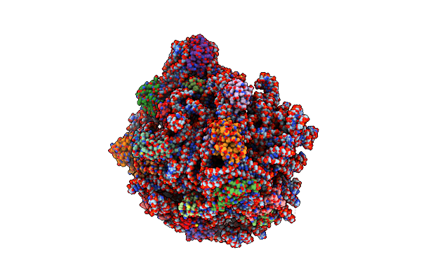
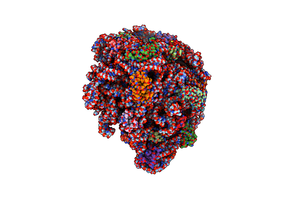
50S Ribosomal Subunit From Staphylococcus Aureus Containing Double Mutation In Ul3 Imparting Linezolid Resistance
Organism: Staphylococcus aureus
Method: ELECTRON MICROSCOPY Release Date: 2022-07-06 Classification: RIBOSOME |
 |
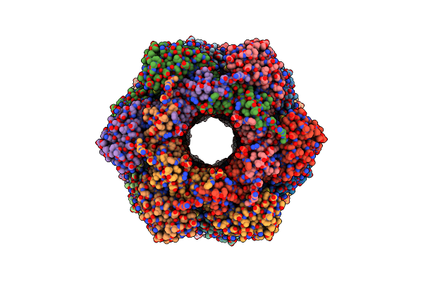
Organism: Klebsiella phage gh-k3
Method: ELECTRON MICROSCOPY Release Date: 2021-08-25 Classification: SUGAR BINDING PROTEIN |
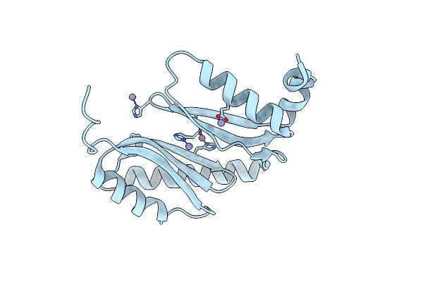 |
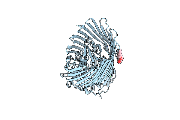
Organism: Acinetobacter baumannii
Method: X-RAY DIFFRACTION Resolution:1.65 Å Release Date: 2021-06-02 Classification: LIPID BINDING PROTEIN Ligands: ZN |
 |
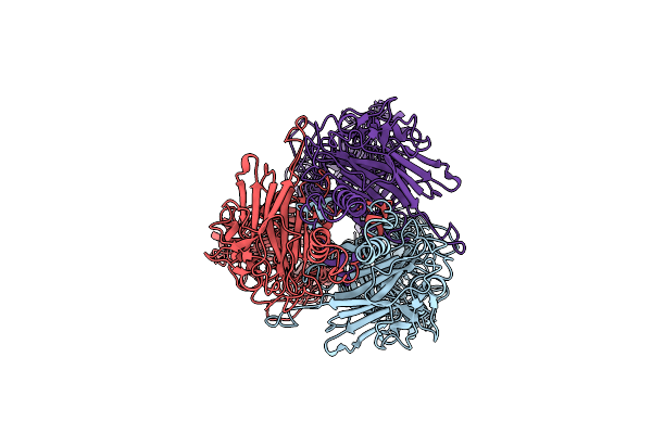
Structure-Function Analyses Of Dual-Bon Domain Protein Dolp Identifies Phospholipid Binding As A New Mechanism For Protein Localisation To The Cell Division Site
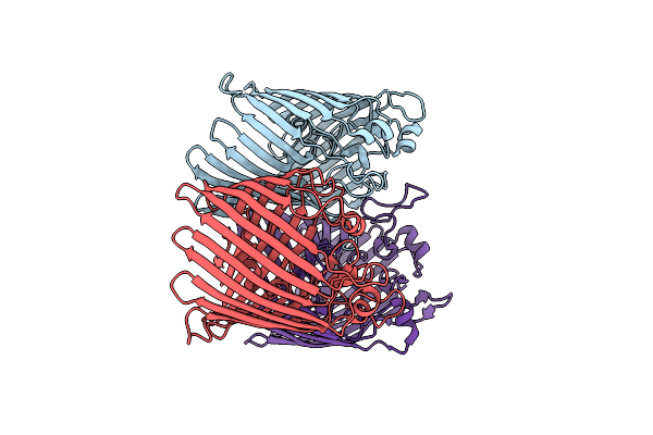
Organism: Escherichia coli (strain k12)
Method: SOLUTION NMR Release Date: 2020-12-30 Classification: PROTEIN BINDING |
 |
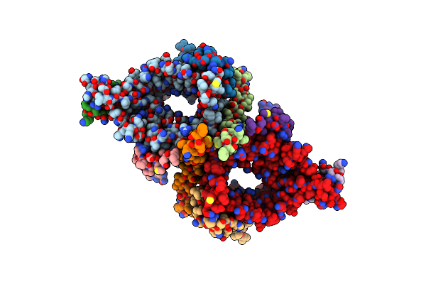
Organism: Bacteriophage sp.
Method: X-RAY DIFFRACTION Resolution:2.60 Å Release Date: 2020-07-01 Classification: VIRAL PROTEIN |
 |
Organism: Bacteriophage sp.
Method: ELECTRON MICROSCOPY Release Date: 2020-07-01 Classification: VIRUS |
 |
Organism: Bacteriophage sp.
Method: ELECTRON MICROSCOPY Release Date: 2020-07-01 Classification: VIRAL PROTEIN |
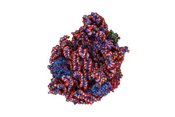 |
Structure Of The 50S Subunit Of The Ribosome From Methicillin Resistant Staphylococcus Aureus In Complex With The Antibiotic, Contezolid
Organism: Staphylococcus aureus
Method: ELECTRON MICROSCOPY Release Date: 2020-06-03 Classification: RIBOSOME Ligands: ZC0 |
 |
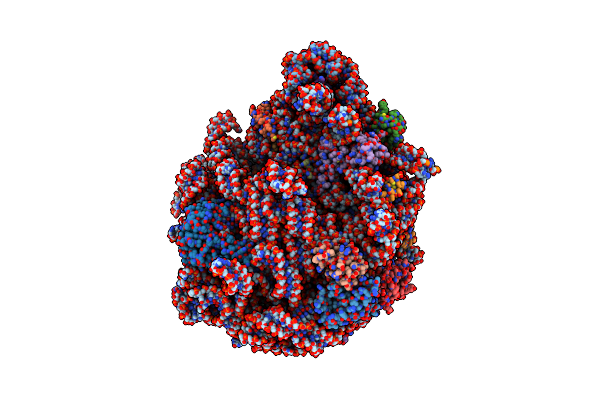
Structure Of The 50S Subunit Of The Ribosome From Methicillin Resistant Staphylococcus Aureus In Complex With The Antibiotic, Radezolid
Organism: Staphylococcus aureus
Method: ELECTRON MICROSCOPY Release Date: 2020-06-03 Classification: RIBOSOME Ligands: RD8 |
 |
Structure Of The 50S Subunit Of The Ribosome From Methicillin Resistant Staphylococcus Aureus In Complex With The Antibiotic, Tedizolid
Organism: Staphylococcus aureus
Method: ELECTRON MICROSCOPY Release Date: 2020-06-03 Classification: RIBOSOME Ligands: U7V |
 |
Structure Of The 50S Subunit Of The Ribosome From Methicillin Resistant Staphylococcus Aureus In Complex With An Isomer Of The Tedizolid
Organism: Staphylococcus aureus
Method: ELECTRON MICROSCOPY Release Date: 2020-06-03 Classification: RIBOSOME Ligands: U7Y |
 |
Organism: Escherichia coli bw25113
Method: X-RAY DIFFRACTION Resolution:2.96 Å Release Date: 2020-05-06 Classification: TRANSPORT PROTEIN Ligands: BOG, CA |
 |
Organism: Klebsiella pneumoniae
Method: X-RAY DIFFRACTION Resolution:2.60 Å Release Date: 2020-04-01 Classification: MEMBRANE PROTEIN |
 |
Organism: Saccharomyces cerevisiae s288c
Method: ELECTRON MICROSCOPY Release Date: 2019-10-16 Classification: TRANSLOCASE Ligands: 46E |
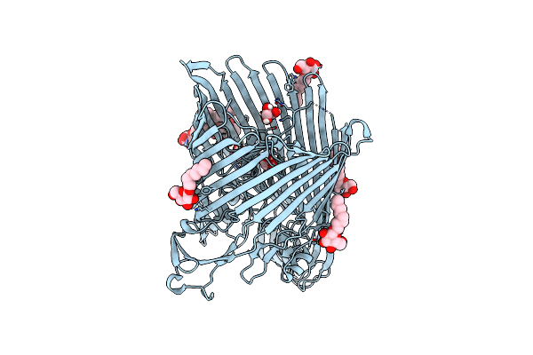 |
The Crystal Structure Of The Outer Membrane Transporter Yddb From Escherichia Coli
Organism: Escherichia coli
Method: X-RAY DIFFRACTION Resolution:2.40 Å Release Date: 2019-10-02 Classification: TRANSPORT PROTEIN Ligands: BOG, MG, GOL |
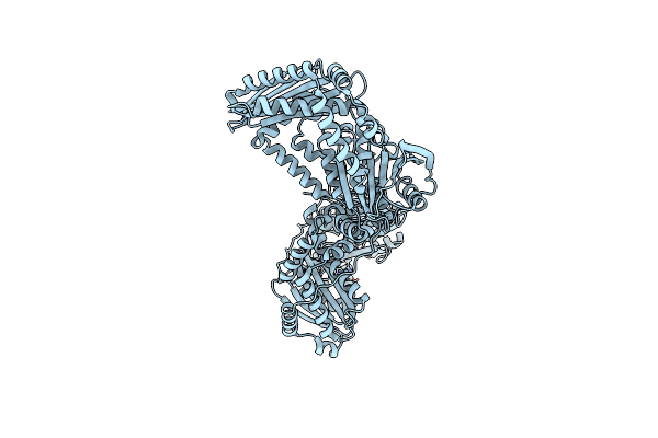 |
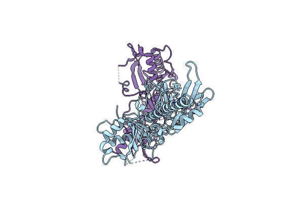
The Crystal Structure Of The Periplasmic Protease Pqql From Escherichia Coli
Organism: Escherichia coli (strain k12)
Method: X-RAY DIFFRACTION Resolution:2.60 Å Release Date: 2019-10-02 Classification: HYDROLASE Ligands: ZN, CL |
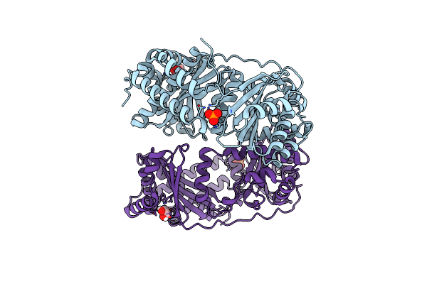 |
The Crystal Structure Of The First Half Of The Periplasmic Protease Pqql From Escherichia Coli
Organism: Escherichia coli (strain k12)
Method: X-RAY DIFFRACTION Resolution:2.00 Å Release Date: 2019-10-02 Classification: HYDROLASE Ligands: GOL, PO4, CL |
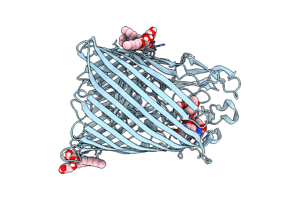 |
The Crystal Structure Of Fhue From E. Coli In Complex With Its Substrate Coprogen
Organism: Escherichia coli bw25113
Method: X-RAY DIFFRACTION Resolution:2.00 Å Release Date: 2019-04-10 Classification: TRANSPORT PROTEIN Ligands: BOG, HWS |
 |
Structure Of The 50S Ribosomal Subunit From Methicillin Resistant Staphylococcus Aureus In Complex With The Oxazolidinone Antibiotic Lzd-5
Organism: Staphylococcus aureus
Method: ELECTRON MICROSCOPY Release Date: 2019-03-20 Classification: Ribosome/Antibiotic Ligands: G6V |