
Search Count: 4
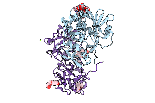 |
The Structure Of The Monoclinic Crystal Form Of The Type Ii Ribosome Inactivating Protein From Winter Aconite Eranthis Hyemalis.
Organism: Eranthis hyemalis
Method: X-RAY DIFFRACTION Resolution:1.50 Å Release Date: 2026-01-21 Classification: TOXIN Ligands: NAG, FUC, PG4, MG |
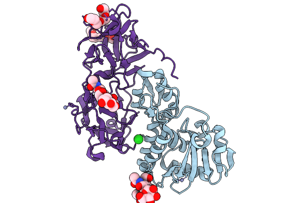 |
The Structure Of The Orthorhombic Crystal Form Of The Type Ii Ribosome Inactivating Protein From Winter Aconite Eranthis Hyemalis.
Organism: Eranthis hyemalis
Method: X-RAY DIFFRACTION Resolution:1.80 Å Release Date: 2026-01-21 Classification: TOXIN Ligands: NAG, MAN, FUC, ZN, CL, PG4, AH2 |
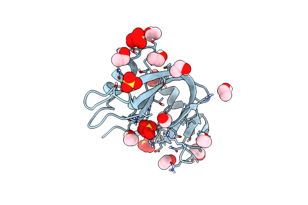 |
Ultra High Resolution X-Ray Structure Of Orthorhombic Bovine Pancreatic Ribonuclease At 100K
Organism: Bos taurus
Method: X-RAY DIFFRACTION Resolution:0.85 Å Release Date: 2022-07-27 Classification: HYDROLASE Ligands: EOH, SO4 |
 |
Organism: Homo sapiens
Method: X-RAY DIFFRACTION Resolution:0.95 Å Release Date: 2016-10-12 Classification: IMMUNE SYSTEM Ligands: ZN, ACT, POL |