
Search Count: 13
 |
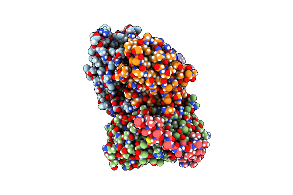
Organism: Homo sapiens, Mus musculus
Method: ELECTRON MICROSCOPY Release Date: 2023-06-14 Classification: MEMBRANE PROTEIN Ligands: NAG, CA, ZN, BAT, Y01 |
 |
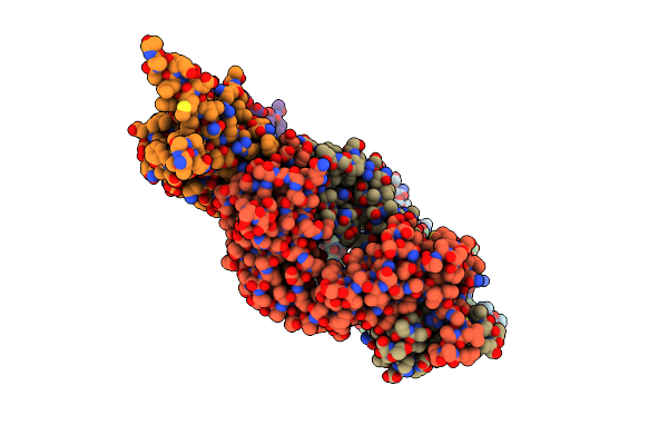
Organism: Homo sapiens, Lama glama
Method: ELECTRON MICROSCOPY Release Date: 2023-05-10 Classification: SIGNALING PROTEIN Ligands: LPC |
 |
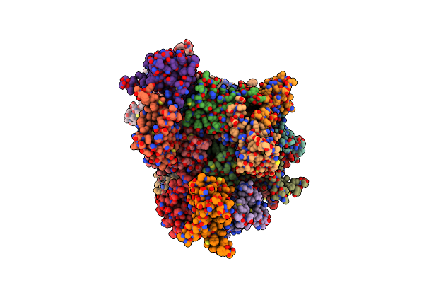
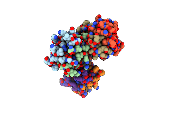
Crystal Structure Of Tspan15 Large Extracellular Loop (Tspan15 Lel) In Complex With 1C12 Fab
Organism: Mus musculus, Homo sapiens
Method: X-RAY DIFFRACTION Resolution:3.60 Å Release Date: 2021-11-03 Classification: CELL ADHESION |
 |
Organism: Homo sapiens
Method: X-RAY DIFFRACTION Resolution:2.52 Å Release Date: 2021-11-03 Classification: CELL ADHESION |
 |
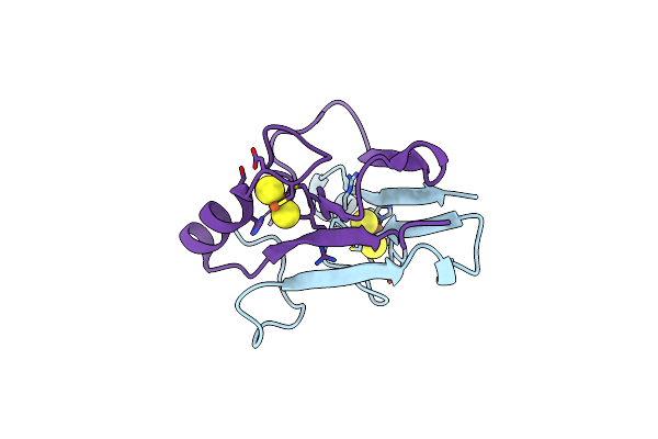
Crystal Structure Of Mutant B1 Immunoglobulin-Binding Domain Of Streptococcal Protein G (T16F, T18A, V21E, T25L, K28Y, V29I, K31R, Q32H, Y33L, N35K, D36H, N37Q)
Organism: Streptococcus
Method: X-RAY DIFFRACTION Resolution:1.40 Å Release Date: 2019-01-23 Classification: DE NOVO PROTEIN Ligands: ZN, CL |
 |
Crystal Structure Of B1 Immunoglobulin-Binding Domain Of Streptococcal Protein G (T16F, T18A, V21H, T25H, K28Y, V29I, K31R, Q32A, Y33L, N35K, D36A, N37Q)
Organism: Streptococcus
Method: X-RAY DIFFRACTION Resolution:1.40 Å Release Date: 2019-01-23 Classification: METAL BINDING PROTEIN Ligands: ZN, ACT, NA, CL, PO4, DPO |
 |
Crystal Structure Of De Novo Designed Metal-Controlled Dimer Of Mutant B1 Immunoglobulin-Binding Domain Of Streptococcal Protein G (L12H, T16L, V29H, Y33H, N37L)-Zinc
Organism: Streptococcus
Method: X-RAY DIFFRACTION Resolution:1.50 Å Release Date: 2019-01-23 Classification: METAL BINDING PROTEIN Ligands: ZN, CL |
 |
Crystal Structure Of De Novo Designed Metal-Controlled Dimer Of Mutant B1 Immunoglobulin-Binding Domain Of Streptococcal Protein G (L12H, T16L, V29H, Y33H, N37L)-Apo
Organism: Streptococcus
Method: X-RAY DIFFRACTION Resolution:1.70 Å Release Date: 2019-01-23 Classification: DE NOVO PROTEIN Ligands: MG, NA |
 |
Crystal Structure Of De Novo Designed Metal-Controlled Dimer Of B1 Immunoglobulin-Binding Domain Of Streptococcal Protein G (L12H, E15V, T16L, T18I, V29H, Y33H, N37L)-Zinc
Organism: Streptococcus
Method: X-RAY DIFFRACTION Resolution:1.34 Å Release Date: 2019-01-23 Classification: DE NOVO PROTEIN Ligands: ZN, CL, GOL, NA |
 |
Crystal Structure Of De Novo Designed Metal-Controlled Dimer Of Mutant B1 Immunoglobulin-Binding Domain Of Streptococcal Protein G (L12H, E15V, T16L, T18I, V29H, Y33H, N37L)-Apo
Organism: Streptococcus
Method: X-RAY DIFFRACTION Resolution:2.30 Å Release Date: 2019-01-23 Classification: METAL BINDING PROTEIN Ligands: MG |
 |
Organism: Homo sapiens
Method: X-RAY DIFFRACTION Resolution:1.90 Å Release Date: 2017-12-20 Classification: METAL BINDING PROTEIN Ligands: FES |
 |
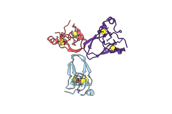
The 1.55A Crystal Structure Of Naf1 (Miner1): The Redox-Active 2Fe-2S Protein
Organism: Homo sapiens
Method: X-RAY DIFFRACTION Resolution:1.65 Å Release Date: 2014-07-02 Classification: METAL BINDING PROTEIN Ligands: FES |
 |
Organism: Homo sapiens
Method: X-RAY DIFFRACTION Resolution:1.58 Å Release Date: 2014-07-02 Classification: METAL BINDING PROTEIN Ligands: FES |