
Search Count: 16
 |
Organism: Homo sapiens, Oplophorus gracilirostris, Rattus norvegicus, Synthetic construct
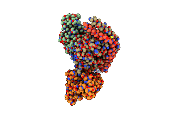
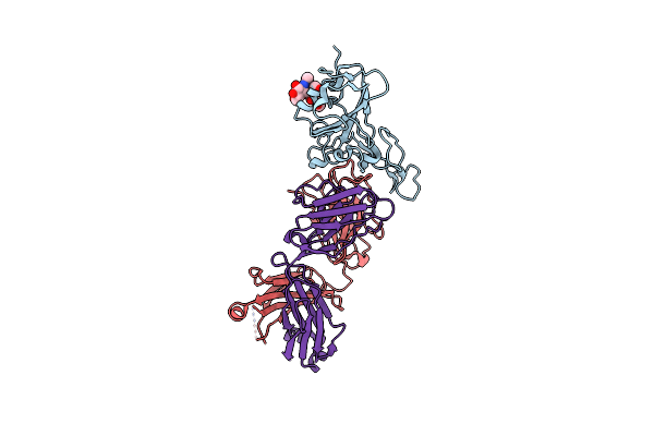
Method: ELECTRON MICROSCOPY Release Date: 2024-07-10 Classification: MEMBRANE PROTEIN/IMMUNE SYSTEM |
 |
Organism: Homo sapiens, Rattus norvegicus, Bos taurus, Oplophorus gracilirostris, Synthetic construct
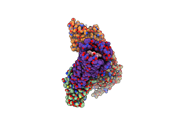
Method: ELECTRON MICROSCOPY Release Date: 2024-07-10 Classification: MEMBRANE PROTEIN/IMMUNE SYSTEM |
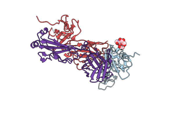 |
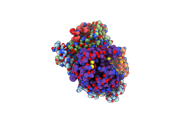
Crystal Structure Of Sars-Cov-2 Receptor Binding Domain In Complex With Sars-Cov-2 Reactive Human Antibody Cr3022
Organism: Homo sapiens, Severe acute respiratory syndrome coronavirus 2
Method: X-RAY DIFFRACTION Resolution:4.22 Å Release Date: 2023-12-13 Classification: VIRAL PROTEIN/IMMUNE SYSTEM |
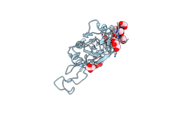 |
Organism: Severe acute respiratory syndrome coronavirus 2
Method: X-RAY DIFFRACTION Resolution:1.95 Å Release Date: 2023-12-13 Classification: VIRAL PROTEIN Ligands: NAG, FUC, GOL |
 |
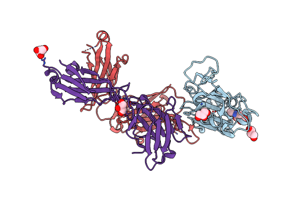
Crystal Structure Of Antibody Wrair-2123 In Complex With Sars-Cov-2 Receptor Binding Domain
Organism: Severe acute respiratory syndrome coronavirus 2, Homo sapiens
Method: X-RAY DIFFRACTION Resolution:3.50 Å Release Date: 2023-12-13 Classification: VIRAL PROTEIN/IMMUNE SYSTEM |
 |
Crystal Structure Of Antibody Wrair-2134 In Complex With Sars-Cov-2 Receptor Binding Domain
Organism: Homo sapiens, Severe acute respiratory syndrome coronavirus 2
Method: X-RAY DIFFRACTION Resolution:3.16 Å Release Date: 2023-06-28 Classification: VIRAL PROTEIN/IMMUNE SYSTEM Ligands: NAG |
 |
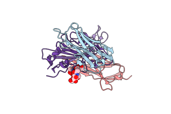
Crystal Structure Of Antibody Ab246 In Complex With Sars-Cov-2 Receptor Binding Domain
Organism: Severe acute respiratory syndrome coronavirus 2, Homo sapiens
Method: X-RAY DIFFRACTION Resolution:2.29 Å Release Date: 2023-03-15 Classification: VIRAL PROTEIN/IMMUNE SYSTEM Ligands: NAG, GOL, CL |
 |
Crystal Structure Of Sars Cov-2 Spike Receptor Binding Domain In Complex With Shark Neutralizing Vnars Shab01 And Shab02
Organism: Ginglymostoma cirratum, Severe acute respiratory syndrome coronavirus 2
Method: X-RAY DIFFRACTION Resolution:2.52 Å Release Date: 2022-11-23 Classification: IMMUNE SYSTEM/VIRAL PROTEIN Ligands: GOL |
 |
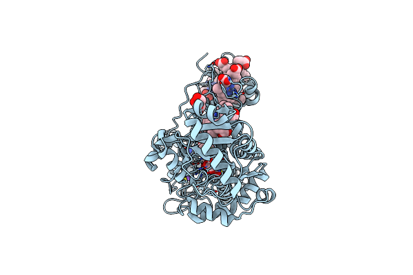
Cryo-Em Structure Of The Partial Agonist Salbutamol-Bound Beta2 Adrenergic Receptor-Gs Protein Complex.
Organism: Homo sapiens, Bos taurus, Lama glama
Method: ELECTRON MICROSCOPY Release Date: 2020-12-16 Classification: SIGNALING PROTEIN Ligands: 68H |
 |
Cryo-Em Structure Of The Full Agonist Isoprenaline-Bound Beta2 Adrenergic Receptor-Gs Protein Complex.
Organism: Homo sapiens, Bos taurus, Lama glama
Method: ELECTRON MICROSCOPY Release Date: 2020-12-16 Classification: SIGNALING PROTEIN Ligands: 5FW |
 |
Crystal Structure Of The Cedar Henipavirus Attachment G Glycoprotein Global Domain
Organism: Cedar virus
Method: X-RAY DIFFRACTION Resolution:3.28 Å Release Date: 2019-09-25 Classification: VIRAL PROTEIN Ligands: NAG |
 |
Crystal Structure Of The Cedar Henipavirus Attachment G Glycoprotein Globular Domain In Complex With The Receptor Ephrin-B1
Organism: Cedar virus, Mus musculus
Method: X-RAY DIFFRACTION Resolution:3.49 Å Release Date: 2019-09-25 Classification: VIRAL PROTEIN Ligands: NAG |
 |
Crystal Structure Of The Cedar Henipavirus Attachment G Glycoprotein Globular Domain In Complex With The Receptor Ephrin-B2
Organism: Cedar virus, Homo sapiens
Method: X-RAY DIFFRACTION Resolution:2.84 Å Release Date: 2019-09-25 Classification: VIRAL PROTEIN Ligands: NAG |
 |
Organism: Mus musculus
Method: X-RAY DIFFRACTION Resolution:2.49 Å Release Date: 2014-02-19 Classification: IMMUNE SYSTEM |
 |
Flavocytochrome C3 From Shewanella Frigidimarina Histidine 365 Mutated To Alanine
Organism: Shewanella frigidimarina
Method: X-RAY DIFFRACTION Resolution:1.80 Å Release Date: 2000-09-18 Classification: OXIDOREDUCTASE Ligands: HEC, FAD, FUM, NA, GOL |
 |
Organism: Shewanella frigidimarina
Method: X-RAY DIFFRACTION Resolution:1.80 Å Release Date: 1999-11-21 Classification: OXIDOREDUCTASE Ligands: HEC, FAD, TEO, NA, GOL |