Search Count: 336
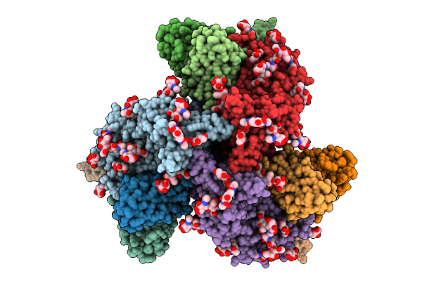 |
Organism: Human immunodeficiency virus 1, Homo sapiens
Method: ELECTRON MICROSCOPY Release Date: 2026-01-21 Classification: IMMUNE SYSTEM Ligands: NAG |
 |
Organism: Homo sapiens, Human immunodeficiency virus 1
Method: ELECTRON MICROSCOPY Release Date: 2026-01-21 Classification: IMMUNE SYSTEM Ligands: NAG |
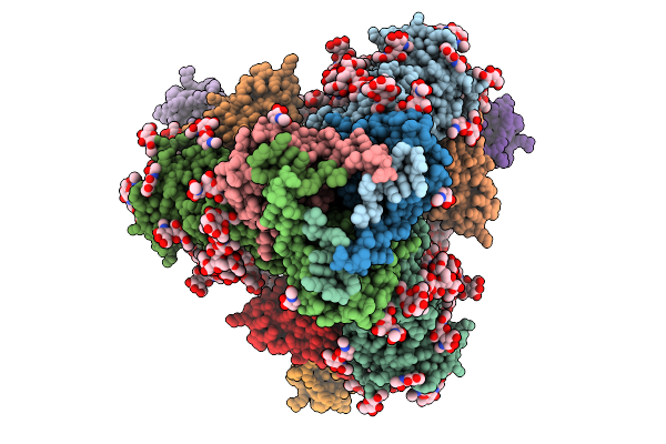 |
Organism: Human immunodeficiency virus 1, Homo sapiens
Method: ELECTRON MICROSCOPY Release Date: 2026-01-21 Classification: IMMUNE SYSTEM Ligands: NAG |
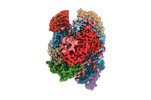 |
Organism: Human immunodeficiency virus 1, Homo sapiens
Method: ELECTRON MICROSCOPY Release Date: 2026-01-21 Classification: IMMUNE SYSTEM Ligands: NAG |
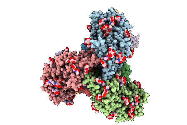 |
Organism: Human immunodeficiency virus 1, Homo sapiens
Method: ELECTRON MICROSCOPY Release Date: 2026-01-21 Classification: IMMUNE SYSTEM Ligands: NAG |
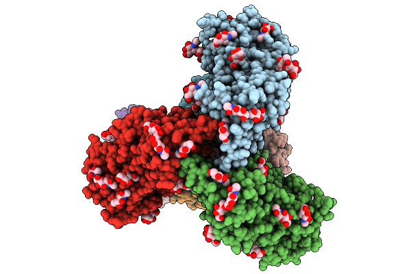 |
Organism: Human immunodeficiency virus 1, Homo sapiens
Method: ELECTRON MICROSCOPY Release Date: 2026-01-21 Classification: IMMUNE SYSTEM Ligands: NAG |
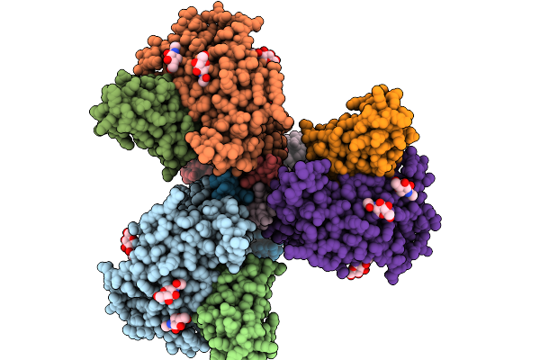 |
Organism: Homo sapiens, Human immunodeficiency virus 1
Method: ELECTRON MICROSCOPY Release Date: 2026-01-21 Classification: IMMUNE SYSTEM Ligands: NAG |
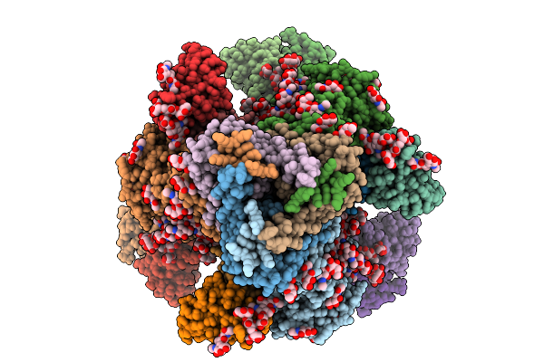 |
Organism: Homo sapiens, Human immunodeficiency virus 1
Method: ELECTRON MICROSCOPY Release Date: 2026-01-21 Classification: IMMUNE SYSTEM Ligands: NAG |
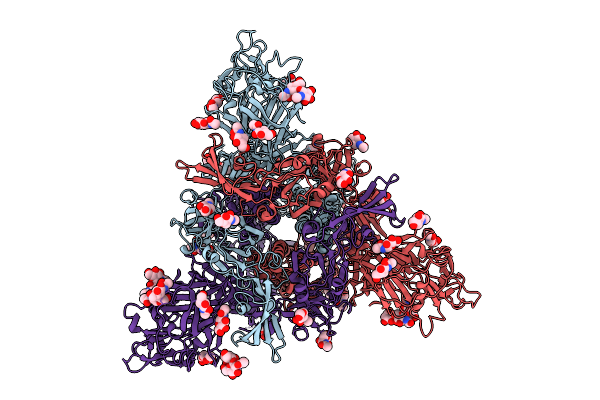 |
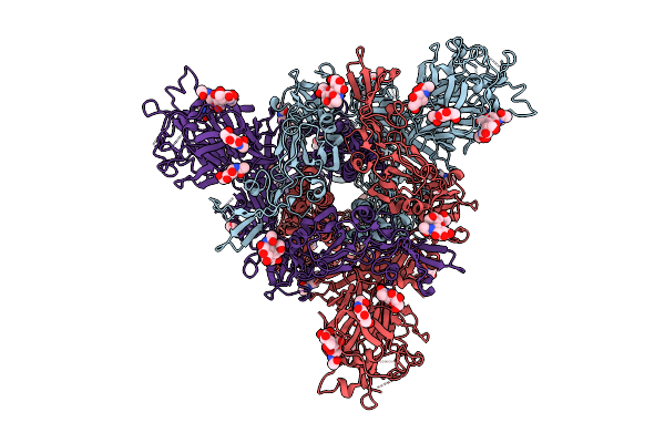
Organism: Bttp-betacov/gx2012
Method: ELECTRON MICROSCOPY Release Date: 2025-12-24 Classification: VIRAL PROTEIN Ligands: NAG |
 |
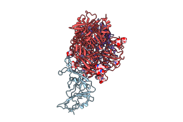
Organism: Middle east respiratory syndrome-related coronavirus
Method: ELECTRON MICROSCOPY Release Date: 2025-12-24 Classification: VIRAL PROTEIN Ligands: NAG |
 |
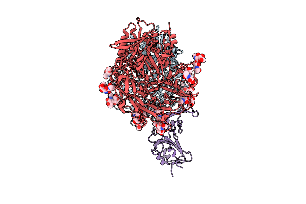
Organism: Middle east respiratory syndrome-related coronavirus, Homo sapiens
Method: ELECTRON MICROSCOPY Release Date: 2025-12-24 Classification: VIRAL PROTEIN/HYDROLASE Ligands: NAG |
 |
Organism: Homo sapiens, Bttp-betacov/gx2012
Method: ELECTRON MICROSCOPY Release Date: 2025-12-24 Classification: VIRAL PROTEIN/HYDROLASE Ligands: NAG |
 |
Organism: Human immunodeficiency virus 1, Homo sapiens
Method: ELECTRON MICROSCOPY Release Date: 2025-12-17 Classification: VIRAL PROTEIN/IMMUNE SYSTEM Ligands: NAG |
 |
Organism: Human immunodeficiency virus 1, Homo sapiens
Method: ELECTRON MICROSCOPY Release Date: 2025-12-17 Classification: VIRAL PROTEIN/IMMUNE SYSTEM Ligands: NAG |
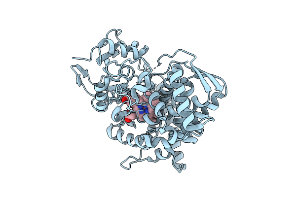 |
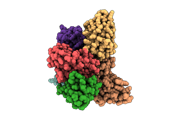
Organism: Novosphingobium sp. 28-62-57
Method: X-RAY DIFFRACTION Resolution:2.10 Å Release Date: 2025-12-17 Classification: OXIDOREDUCTASE Ligands: HEM |
 |
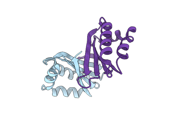
Organism: Homo sapiens, Plasmodium falciparum
Method: ELECTRON MICROSCOPY Release Date: 2025-12-10 Classification: IMMUNE SYSTEM/DE NOVO PROTEIN |
 |
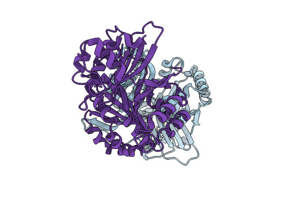
An Antibiotic Biosynthesis Monooxygenase Family Protein From Streptomyces Sp. Ma37
Organism: Streptomyces sp. ma37
Method: X-RAY DIFFRACTION Resolution:1.65 Å Release Date: 2025-11-26 Classification: ANTIBIOTIC |
 |
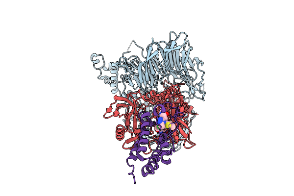
Crystal Structure Of A Polyketide Abm/Scha-Like Domain-Containing Protein Whie-Orfi From Streptomyces Coelicolor
Organism: Streptomyces coelicolor
Method: X-RAY DIFFRACTION Resolution:2.30 Å Release Date: 2025-11-26 Classification: ANTIBIOTIC |
 |
Ternary Protac-Mediated Complex Consisting Of Cereblon, Ddb1 And Brd4-Bd1, Non-Covalently Linked By Jq1-Acn
Organism: Homo sapiens
Method: ELECTRON MICROSCOPY Resolution:2.66 Å Release Date: 2025-10-29 Classification: LIGASE Ligands: ZN, A1JM3 |
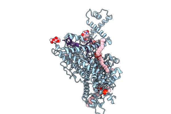 |
Vitamin K-Dependent Gamma-Carboxylase With Osteocalcin (Mutant) And Vitamin K Hydroquinone And Calcium
Organism: Homo sapiens
Method: ELECTRON MICROSCOPY Release Date: 2025-10-08 Classification: TRANSFERASE Ligands: NAG, 6PL, A1AVC |

