
Search Count: 17
 |
Organism: Klebsiella phage vlc6
Method: X-RAY DIFFRACTION Resolution:1.38 Å Release Date: 2024-02-21 Classification: HYDROLASE Ligands: TRS, GOL, BCN |
 |
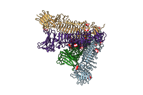
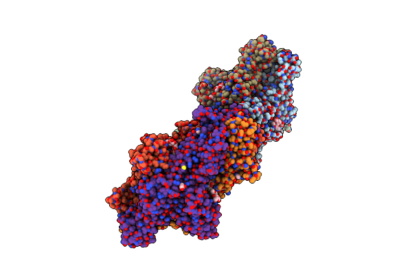
Crystal Structure Of Trimeric K2-2 Tsp In Complex With Tetrasaccharide And Octasaccharide
Organism: Klebsiella phage vlc6
Method: X-RAY DIFFRACTION Resolution:1.58 Å Release Date: 2024-02-21 Classification: HYDROLASE Ligands: ACE, EDO |
 |
Organism: Klebsiella phage vlc6
Method: X-RAY DIFFRACTION Resolution:2.17 Å Release Date: 2024-02-21 Classification: HYDROLASE Ligands: GOL |
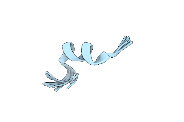 |
Organism: Illicium verum
Method: SOLUTION NMR Release Date: 2023-08-16 Classification: ANTIMICROBIAL PROTEIN |
 |
Organism: Illicium anisatum
Method: SOLUTION NMR Release Date: 2022-11-09 Classification: ANTIMICROBIAL PROTEIN |
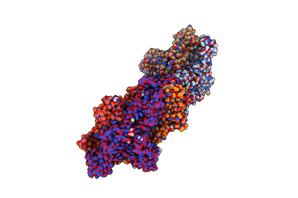 |
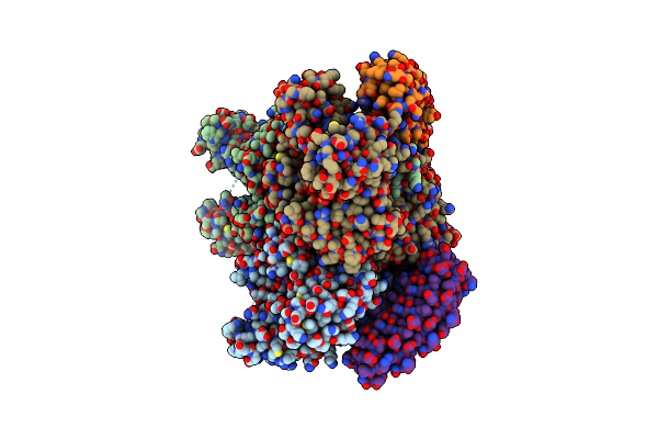
Crystal Structure Of Klebsiella Pneumoniae K1 Capsule-Specific Polysaccharide Lyase In A P1 Crystal Form
Organism: Klebsiella phage ntuh-k2044-k1-1
Method: X-RAY DIFFRACTION Resolution:1.48 Å Release Date: 2022-05-18 Classification: VIRAL PROTEIN Ligands: LMR, IMD, GOL |
 |
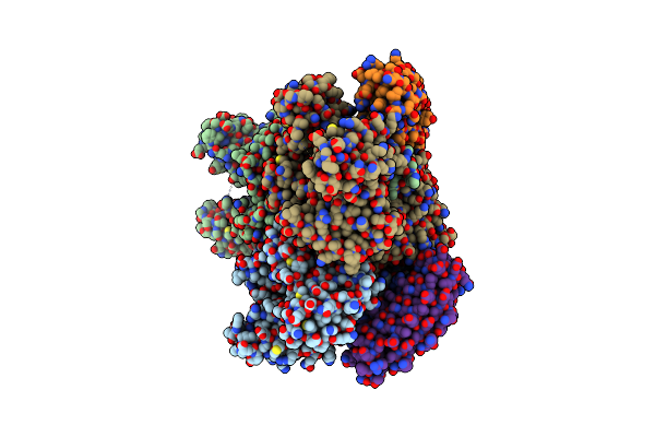
Crystal Structure Of Klebsiella Pneumoniae K1 Capsule-Specific Polysaccharide Lyase In A C2 Crystal Form
Organism: Klebsiella phage ntuh-k2044-k1-1
Method: X-RAY DIFFRACTION Resolution:2.78 Å Release Date: 2022-05-18 Classification: VIRAL PROTEIN Ligands: CIT, CO3 |
 |
Crystal Structure Of Klebsiella Pneumoniae K1 Capsule-Specific Polysaccharide Lyase In Complex With Products
Organism: Klebsiella phage ntuh-k2044-k1-1
Method: X-RAY DIFFRACTION Resolution:1.46 Å Release Date: 2022-05-18 Classification: VIRAL PROTEIN Ligands: IMD, GOL, 98X |
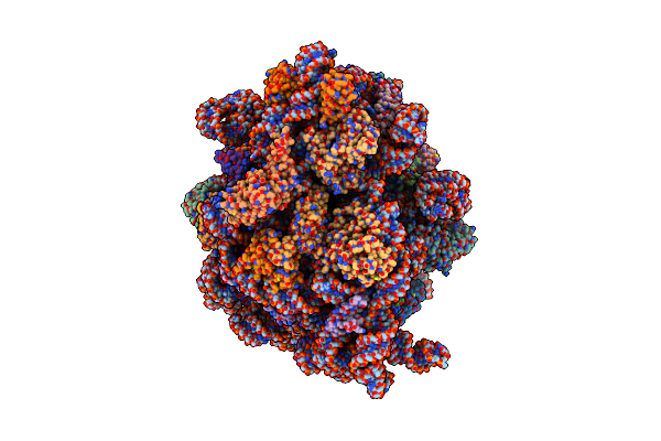 |
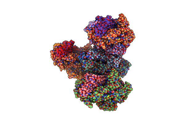
Structure Of Trans-Translation Inhibitor Bound To E. Coli 70S Ribosome With P Site Trna
Organism: Escherichia coli
Method: ELECTRON MICROSCOPY Release Date: 2021-02-10 Classification: RIBOSOME Ligands: KKL |
 |
Organism: Escherichia coli (strain k12), Ribosome display vector prdv
Method: X-RAY DIFFRACTION Resolution:2.30 Å Release Date: 2016-04-06 Classification: TRANSPORT PROTEIN |
 |
Organism: Escherichia coli k-12, Synthetic construct
Method: X-RAY DIFFRACTION Resolution:2.20 Å Release Date: 2016-04-06 Classification: TRANSPORT PROTEIN Ligands: 5QG |
 |
Organism: Escherichia coli (strain k12), Escherichia coli k-12, Synthetic construct
Method: X-RAY DIFFRACTION Resolution:1.90 Å Release Date: 2016-04-06 Classification: TRANSPORT PROTEIN Ligands: 5QF |
 |
Organism: Escherichia coli k-12, Synthetic construct
Method: X-RAY DIFFRACTION Resolution:1.80 Å Release Date: 2016-04-06 Classification: TRANSPORT PROTEIN Ligands: 5QE |
 |
Organism: Escherichia coli k-12, Synthetic construct
Method: X-RAY DIFFRACTION Resolution:2.30 Å Release Date: 2016-04-06 Classification: TRANSPORT PROTEIN Ligands: MBX |
 |
Organism: Escherichia coli k-12, Synthetic construct
Method: X-RAY DIFFRACTION Resolution:2.80 Å Release Date: 2016-04-06 Classification: TRANSPORT PROTEIN Ligands: RHQ |
 |
Organism: Escherichia coli k-12, Synthetic construct
Method: X-RAY DIFFRACTION Resolution:2.50 Å Release Date: 2016-04-06 Classification: TRANSPORT PROTEIN Ligands: MIY, PEG |
 |
Organism: Homo sapiens
Method: X-RAY DIFFRACTION Resolution:2.10 Å Release Date: 1995-02-07 Classification: BINDING PROTEIN |